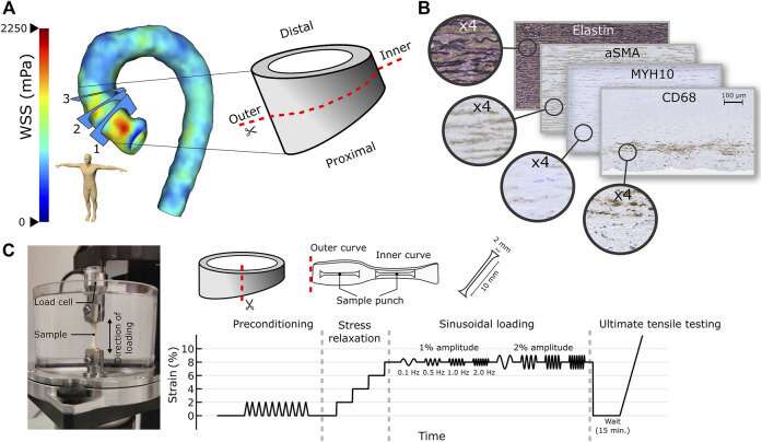
(A) A WSS map of dilated AA along with the three analysis planes and the extracted aortic sample subjected to histological (distal part) and biomechanical (proximal part) measurements are presented. (B) Media degeneration scoring was performed together with histological stainings to detect elastin, smooth muscle cells (α-SMA, MYH10), and inflammatory cells (CD68, CD3) from the inner and outer curves of the AA. (C) Sample processing in the biomechanical measurements along with the measurement system and protocol. Credit: Frontiers in Physiology (2022). DOI: 10.3389/fphys.2022.934941
Prediction of aortic dissection or rupture of the thoracic aortic aneurysm is a challenge, and more accurate identification of high-risk patients and timing of repair surgery are required. A new collaborative study by the University of Eastern Finland and Kuopio University Hospital indicates that magnetic resonance imaging-derived wall shear stress values predict pathological changes in the aortic wall in patients with ascending aortic dilatation. The findings could be used for the identification of high-risk patients in the future.
In ascending aortic dilatation, the aorta progressively dilates due to the weakening of the aortic wall structure leading to the formation of an aneurysm. This can lead to severe complications, such as aortic dissection or rupture. The main risk factors for ascending aortic dilatation are the bicuspid aortic valve and certain genetic factors or syndromes.
Besides surgical intervention, no effective treatment exists. Typically, patients are asymptomatic, and an aneurysm is discovered incidentally, or only when a life-threatening complication occurs. After diagnosis, aortic dilatation is monitored regularly by computed tomography or magnetic resonance imaging.
Besides genetics, aortic vascular flow has been suggested to induce changes in the aortic wall structure and to contribute to disease progression. The new study from the University of Eastern Finland and Kuopio University Hospital shows for the first time the relation between wall shear stress, aortic wall strength, and site-specific cell composition in patients having ascending aortic dilatation.
The results demonstrate that altered wall shear stress in patients regulates biomechanical properties, aortic degeneration, and cell-driven mechanisms. The study identifies a new molecular marker, MYH10, as an indicator of pathological changes in the aortic wall. These findings may be used for the identification of high-risk patients in the future.
"Our research aim is to identify novel molecular targets for aortic dilatation that could be used to develop their therapeutics and diagnostics," says Academy Research Fellow Johanna Laakkonen of the University of Eastern Finland.
"We aim to identify patients with a high risk of aortic dilatation or rupture and improve the follow-up of these patients, thus reducing the risk for life-threatening complications," says Professor Marja Hedman of the University of Eastern Finland and Kuopio University Hospital.
The study, openly accessible in Frontiers in Physiology, was carried out by the Vascular Biology research group of Academy Research Fellow, Adjunct Professor Johanna Laakkonen at the A.I. Virtanen Institute for Molecular Sciences, and by the research group of Professor Marja Hedman at the School of Medicine, Institute of Clinical Medicine and Kuopio University Hospital Heart Center and Clinical Imaging Center.
More information: Miika Kiema et al, Wall Shear Stress Predicts Media Degeneration and Biomechanical Changes in Thoracic Aorta, Frontiers in Physiology (2022). DOI: 10.3389/fphys.2022.934941
Journal information: Frontiers in Physiology
Provided by University of Eastern Finland


