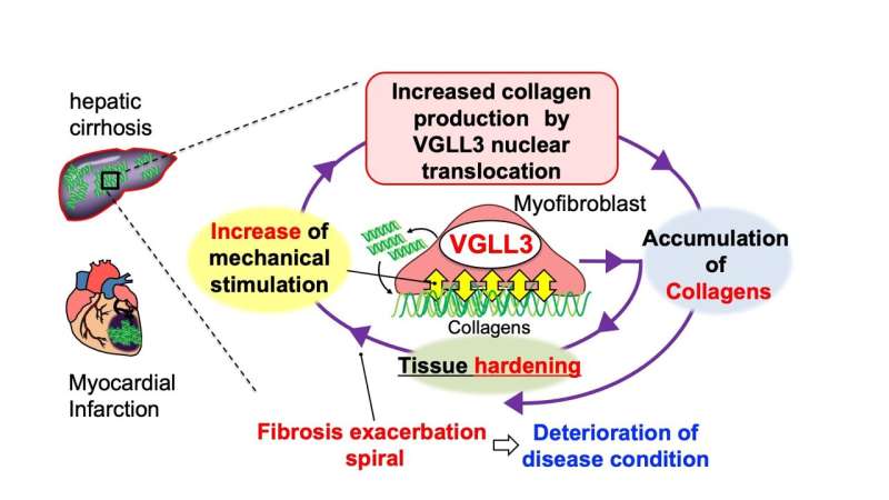
Myofibroblasts are cells that produce collagen and are key in wound repair. Mechanical stimuli of myofibroblast induces VGLL3 to produce more collagen, resulting in hardening of the extracellular matrix and the tissue. The hardening results in an increase in mechanical stimuli that again induces VGLL3 to produce more collagen. The result is a continuing death spiral of fibrosis. Credit: Nakaya Lab/Kyushu University
Researchers from Kyushu University have found how a single mechanosensitive protein induces the process that thickens and scars tissue, known as fibrosis. The protein, called VGLL3, was shown to contribute to fibrosis in multiple organs.
The team hope their findings will lead to new treatments against fibrosis, a pathology that is attributed to 45% of all deaths in industrial nations. Their study was published in Nature Communications.
In response to any injury, the body immediately begins a stream of events. Blood coagulates, the tissue begins to inflame, and the body begins to heal. In some cases that healing comes in the form of scaring and hardening. When an injury is on your skin, it shows up as a visible scar, but what happens when vital organs like your heart or liver are damaged and hardens? If left unchecked, it can lead to loss of mechanics and dangerous consequences.
These changes in tissues are attributed to the extracellular matrix. The extracellular matrix is a web of proteins found in every cell in the body, and acts both like wires on a circuit that allow cells to communicate with each other, and the beams in a building, giving the organs its structure.
Too much extracellular matrix makes the cell, and by extension the organ, tough and inflexible, a condition known as fibrosis. In simple terms, fibrosis is a stiffening of cells and tissue. Its health implications are profound, as it can lead to poor pumping by the heart or cirrhosis in the liver.
"Myofibroblasts are a group of cells that produce collagen, a common extracellular matrix protein. In diseased organs they are seen overproducing collagen. Once myofibroblasts appear in diseased organs, fibrosis proceeds in a snowball fashion," says Michio Nakaya Associate Professor at Kyushu University's Faculty of Pharmaceutical Sciences who led the study. "At the same time, myofibroblasts are responsible for proper wound healing."
To understand how myofibroblasts turn pathological, Nakaya and his colleagues looked at how different physical stimuli changes the expression of genes in these cells. They found consistent changes in the expression of one gene: VGLL3.
Their study showed that after a heart attack, myofibroblasts in both mouse and human hearts express more VGLL3 protein which led to the production of collagen. VGLL3 was also expressed more in fibrotic mouse liver, suggesting it contributes to fibrosis in multiple organs. Conversely, preventing VGLL3 activation in mice led to far less fibrosis in these organs.
"We found that VGLL3 is translocated from the cytoplasm to the nucleus and begins to produce collage in response to mechanical stimuli. In mice without VGLL3, the fibrosis after a heart attack was reduced," noted Nakaya.
The study further showed that the relationship between matrix stiffness and VGLL3 activation becomes a pathological positive feedback loop, in that a stiffer matrix triggers more VGLL3 activation, which triggers the cell to produce more collagen.
Currently, there are only three drugs available for the treatment of fibrosis, and each has its limitations. Considering VGLL3's effects on cell stiffness, Nakaya believes research on treatments should give more attention to this protein.
"In the future, we expect to develop drugs and therapies for fibrosis by targeting VGLL3," concludes Nakaya.
More information: Yuma Horii et al, VGLL3 is a mechanosensitive protein that promotes cardiac fibrosis through liquid–liquid phase separation, Nature Communications (2023). DOI: 10.1038/s41467-023-36189-6
Journal information: Nature Communications
Provided by Kyushu University


