This article has been reviewed according to Science X's editorial process and policies. Editors have highlighted the following attributes while ensuring the content's credibility:
fact-checked
trusted source
proofread
Diagnosing pulmonary hypertension through non-invasive methods

Pulmonary hypertension, or high blood pressure in the lungs, is a common complication of interstitial lung disease, an array of conditions that cause scarring of the lungs. Without treatment, it can be life-threatening. But currently, the only way to diagnose pulmonary hypertension definitively is through an invasive outpatient procedure called right heart catheterization, which measures pressures inside the heart and lungs using a small device inserted through a neck vein.
Because non-invasive tests alone are insufficient to detect pulmonary hypertension, the diagnosis of the condition in the context of interstitial lung disease is challenging. Thus, a team led by Phillip Joseph, MD, assistant professor of medicine (pulmonary) and associate director of the Yale Pulmonary Vascular Disease (PVD) Program, sought to evaluate the effectiveness of a range of variables measured through standard non-invasive tests.
The study identified which variables were the strongest predictors of pulmonary hypertension. It also found a combination of variables that could predict the condition with high accuracy. The team published its findings in Pulmonary Circulation.
"Using our study and combinations of variables, we were able to predict the presence of pulmonary hypertension within interstitial lung disease," says Joseph, who was co-first author. "Using this combination can give us a good pre-procedure probability that someone has pulmonary hypertension, and with more studies, potentially reduce the need for right heart catheterization."
Pulmonary hypertension diagnosis now requires invasive testing
Patients with interstitial lung disease experience inflammation and scarring of the lung tissues which can cause low oxygen levels, shortness of breath, and exercise intolerance. These symptoms can be further exacerbated by pulmonary hypertension. After a patient goes through an initial assessment, if the clinician suspects elevated pressure, the patient undergoes a right heart catheterization to confirm a diagnosis. Then, the clinician can begin to administer necessary medications.
Current non-invasive approaches for detecting pulmonary hypertension are useful screening tools, but lack the specificity needed to officially diagnose the condition. Therefore, right heart catheterization, although invasive, remains the gold standard for diagnosis.
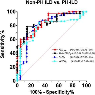
In its latest study, the team enrolled patients who had been previously diagnosed with pulmonary hypertension through right heart catheterization. Then, they evaluated the correlation of several non-invasive measurable variables with a pulmonary hypertension diagnosis. Those measures included as six-minute walk distance, diffusing capacity for carbon monoxide (DLCO) [measured through a pulmonary function test], greater forced vital capacity (FVC) [how much patient can forcibly exhale] to DLCO ratio, right ventricular systolic pressure (RVSP) [measured through echocardiogram], and hemodynamics [pressure measurements].
Non-invasive variables predict pulmonary hypertension
The study revealed that two non-invasive variables in particular—gas exchange-derived pulmonary vascular capacitance [measured during exercise tests] and delta end-tidal carbon dioxide [measures cardiac output]—were the strongest in determining whether interstitial lung disease with pulmonary hypertension was present. Furthermore, using CART statistical analysis, the team found that the combination of gas exchange-derived pulmonary vascular capacitance, estimated right ventricular systolic pressure, and an elevated FVC to DLCO ratio can predict pulmonary hypertension in interstitial lung disease with high sensitivity and specificity.
"We are excited to have this helpful adjunct to our routine assessment," says Danielle Antin-Ozerkis, MD, associate professor of medicine (pulmonary). "If we can only perform catheterization on those who need it and avoid invasive testing in those who don't, that is a win for our patients."
Fortunately for patients with a pulmonary hypertension diagnosis, medications are effective in reducing poor outcomes and improving breathing and exercise tolerance. Because earlier use of medication results in better outcomes, the team hopes its work can help lead to faster and more efficient diagnoses.
More information: Phillip Joseph et al, Noninvasive determinants of pulmonary hypertension in interstitial lung disease, Pulmonary Circulation (2023). DOI: 10.1002/pul2.12197


