May 9, 2023 report
This article has been reviewed according to Science X's editorial process and policies. Editors have highlighted the following attributes while ensuring the content's credibility:
fact-checked
peer-reviewed publication
trusted source
proofread
Glioblastoma found to hijack cognitive neural networks with thrombospondin-1

Researchers from the University of California at San Francisco have examined how glioma-induced neuronal changes influence the brain circuitry underlying cognition and whether these interactions can influence a patient's survival.
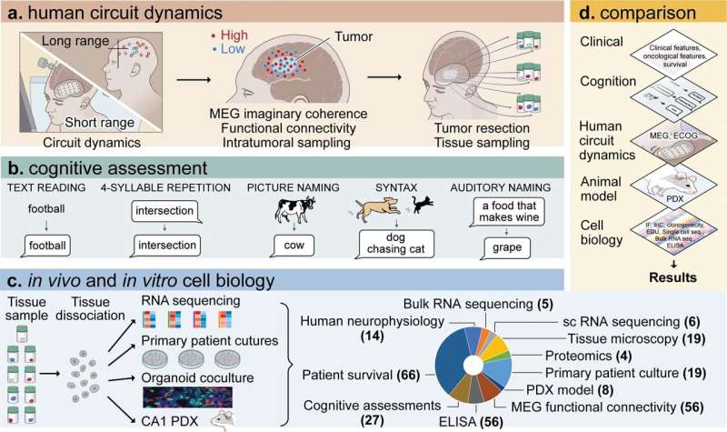
The paper, "Glioblastoma remodeling of human neural circuits decreases survival," published in Nature, finds that gliomas, a type of tumor-forming cell, can alter the functional neural circuits in the brain. This results in activating tumor-infiltrated white matter beyond the usual cortical regions recruited in a healthy brain during language tasks involving lexical retrieval.
Lexical retrieval is the process that takes place in the brain while speaking, from concept to utterance. In the simple task of seeing a picture of a cow and saying the word cow, the brain processes the concept (cow), accesses the appropriate word associated with the image (cow), and the body voices the word "cow." Word loss or replacement occurs when there is interference along the lexical retrieval process.
Several things can affect the lexical retrieval process. For instance, uncommonly used words will have weaker neural reinforcement, making remembering the word more complex than commonly used and synaptically reinforced words. Interference can come about through head trauma that causes inflammation or neurodegeneration, where synaptic connections along the retrieval route are destroyed or remodeled in such a way as to make the connections inoperable.
A patient can have a range of reactions when the retrieval process is disrupted, from not being able to retrieve the word even though the image is familiar—that "tip-of-the-tongue" feeling—or the subject might unknowingly swap the word for something else, like seeing a picture of a cow and saying "key" despite having no confusion about the concept of a cow.
Neural processing of speech can vary by word complexity and frequency of use. Vocalizing less frequently used words requires more intricate coordination than commonly used (high-frequency) words. Researchers determined the decodability of neuronal signals from normal-appearing and glioblastoma-infiltrated cortex using a logistic regression classifier to distinguish between low-frequency and high-frequency word trial conditions.
Patients with glioma often experience lexical retrieval issues, but whether the tumors remodel or destroy the neuronal circuits in the process pathway was unknown.
In experimental conditions, normal brain tissue produced above-chance decoding between low- and high-frequency word trials. By contrast, glioblastoma-infiltrated brain tissue did not decode word trials above chance. This demonstrated to the researchers that glioblastoma infiltration maintains task-specific neuronal responses, the task of accessing a word, yet the tumor-affected brain regions lose the ability to decode complex or low-frequency words.
Using intracranial brain recordings during lexical retrieval language tasks, tumor tissue biopsies, and cell biology experiments, the team found gliomas physically remodeling functional neural circuitry. Task-relevant neural responses activated tumor-inflated cortex areas well beyond the cortical regions usually recruited in the healthy brain. Biopsies from these tumor regions exhibiting high functional connectivity with the rest of the brain were enriched with a specific glioblastoma subpopulation that shows a distinct synaptogenic and neurotrophic phenotype—synaptogenic factor thrombospondin-1 (TSP-1).
TSP-1 is a synaptogenic factor secreted in the healthy brain by astrocytes and is associated with the assembly of neural circuits, including synapse-associated genes and axon pathfinding genes. TSP-1 has long been associated with gliomas and is known to play a role in the mechanisms that control changes to neuronal morphology and brain plasticity. The current research discovered a much more nefarious relationship between TSP-1 and neural networks.
The study found feedback between high-grade gliomas and neural networks was bidirectional, with neuronal activity promoting glioma growth and gliomas increasing neuronal excitability. TSP-1-expressing malignant cells interacted with neuronal signals, exhibiting a synaptogenic, proliferative, invasive and integrative profile. The TSP-1 tumors were effectively hijacking cognitive function, the act of lexicon retrieval, to fuel their growth and create more connections beyond the usual retrieval process to further their spread.
In an additional leg of the study, mice with functional connectivity between the tumor and the rest of the brain had shorter overall survival than those without TSP-1-expressing malignant cells. The degree of functional connectivity between glioblastoma and the normal brain was negatively correlated to both mouse survival and human performance in language tasks.
Inhibition of TSP-1 using the FDA-approved drug gabapentin decreased glioblastoma cell proliferation and network synchrony, suggesting a potential future therapeutic intervention strategy.
More information: Saritha Krishna et al, Glioblastoma remodelling of human neural circuits decreases survival, Nature (2023). DOI: 10.1038/s41586-023-06036-1
© 2023 Science X Network


