This article has been reviewed according to Science X's editorial process and policies. Editors have highlighted the following attributes while ensuring the content's credibility:
fact-checked
proofread
Visualizing heart disease to communicate risk improves patient adherence

Visualizing early heart disease using cardiac imaging helps with patient understanding of risk and adherence to medication and lifestyle change, according to a new study by researchers from the Baker Heart and Diabetes Institute and the Menzies Institute for Medical Research.
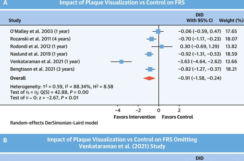
Researchers found patients who viewed calcium or plaque build-up in their arteries using cardiac imaging had improved levels of risk factors like cholesterol and high blood pressure and overall heart risk reduction, the paper in JACC: Cardiovascular Imaging shows.
The study, led by Ph.D. candidate Kristyn Whitmore, involved the analysis of six randomized controlled studies involving more than 7,000 patients and is believed to be the first to determine whether patient visualization of heart images improves the 10-year Framingham risk score and individual risk factors.
Of the six studies, four studies used coronary artery calcium scanning and two used carotid ultrasound to detect subclinical or early atherosclerosis, a thickening of the arteries caused by a build-up of plaque in the inner lining of an artery. All studies used image visualization in the intervention group to communicate heart disease risk.
The analysis demonstrated that not only did cardiac imaging help with diagnosis and risk assessment but importantly, it also assisted in educating and motivating people to engage in risk modification or lifestyle change.
Heart disease is the biggest killer globally and senior author and cardiologist at the Baker Institute, Professor Tom Marwick, says it is critical that we look at novel approaches to recognize and treat heart disease early before significant events like a heart attack or stroke occur.
"This study shows that using pictures as well as conventional risk information is helping to bridge a person's knowledge gap and to improve adherence to both medication and lifestyle change," Professor Marwick says.
Heart disease risk factor identification, risk estimation, and management remain the current standard of care in primary prevention of heart disease, however, Professor Marwick says current risk assessment models have some limitations. In addition to underestimation and overestimation of risk in certain populations, heart risk estimation is a one-off measurement of risk levels at one point in time, so the assessment does not reflect the effect of cumulative exposure of heart disease risk factors over a lifetime.
Heart risk scores have also been reported as being misunderstood or misperceived by people who don't have symptoms. "It can be difficult to get a person who feels completely well to take tablets every day to reduce their cardiac risk," Professor Marwick says. "But once you can show people that the blood vessels are damaged, they realize that it's not about controlling a risk factor that may never cause a problem—it's about treating a disease that has actually started."
A lack of understanding or motivation can lead to people being complacent which can ultimately have catastrophic consequences in the form of a heart attack or stroke, he says.
Professor Marwick says screening tools such as coronary artery calcium scans and carotid ultrasound allow the early identification of subclinical heart disease (before a heart event occurs) in people without symptoms.
He says these tests have the most clinical value in people at intermediate risk who have no prior heart history, by reclassifying them into lower or higher risk groups.
"We need to look closely at how we can better support GPs to promote risk modification and prevention strategies, and patient visualization of early heart damage using cardiac imaging is one tool that warrants further investigation," Professor Marwick says.
"Cardiac imaging is a critical diagnostic tool for cardiologists but this study demonstrates it may also have a much broader use to facilitate communication and understanding. I hope that studies like this may help to justify the wider use of heart imaging like coronary artery calcium scans at a population-based level."
More information: Kristyn Whitmore et al, Impact of Patient Visualization of Cardiovascular Images on Modification of Cardiovascular Risk Factors, JACC: Cardiovascular Imaging (2023). DOI: 10.1016/j.jcmg.2023.03.007


