Radiology & Imaging

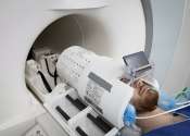
World's most powerful MRI scans first images of human brain

The world's most powerful MRI scanner has delivered its first images of human brains, reaching a new level of precision that is hoped will shed more light on our mysterious minds—and the illnesses that haunt them.

Oncology & Cancer

Myths about PSA tests and prostate cancer screening

The understanding of prostate cancer has significantly evolved in the past 15 years. However, many people—including primary care providers—may not be aware of the current guidelines for prostate cancer screening, says ...

Diseases, Conditions, Syndromes

Researchers develop early detection method for rare Borna virus

Researchers at Augsburg University Medicine have discovered a possible early detection method of the rare Borna virus. Their results have been published in The Lancet. In humans the virus triggers inflammation in the brain ...

page 1 from 2

Magnetic resonance imaging

Magnetic Resonance Imaging (MRI), or nuclear magnetic resonance imaging (NMRI), is primarily a medical imaging technique most commonly used in radiology to visualize the internal structure and function of the body. MRI provides much greater contrast between the different soft tissues of the body than computed tomography (CT) does, making it especially useful in neurological (brain), musculoskeletal, cardiovascular, and oncological (cancer) imaging. Unlike CT, it uses no ionizing radiation, but uses a powerful magnetic field to align the nuclear magnetization of (usually) hydrogen atoms in water in the body. Radio frequency (RF) fields are used to systematically alter the alignment of this magnetization, causing the hydrogen nuclei to produce a rotating magnetic field detectable by the scanner. This signal can be manipulated by additional magnetic fields to build up enough information to construct an image of the body.:36

Magnetic Resonance Imaging is a relatively new technology. The first MR image was published in 1973 and the first cross-sectional image of a living mouse was published in January 1974. The first studies performed on humans were published in 1977. By comparison, the first human X-ray image was taken in 1895.

Magnetic Resonance Imaging was developed from knowledge gained in the study of nuclear magnetic resonance. In its early years the technique was referred to as nuclear magnetic resonance imaging (NMRI). However, as the word nuclear was associated in the public mind with ionizing radiation exposure it is generally now referred to simply as MRI. Scientists still use the term NMRI when discussing non-medical devices operating on the same principles. The term Magnetic Resonance Tomography (MRT) is also sometimes used.

This text uses material from Wikipedia, licensed under CC BY-SA