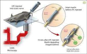
Researchers have recorded how myelin degrades real-time in live mice using a new imaging technique called CARS. In this experiment, the myelin degradation was artificially induced with a compound called lysophosphatidylcholine (LPC). Researchers were able to observe an influx of calcium ions as the myelin began to degrade. This insight could promote early detection of conditions such as multiple sclerosis. Credit: Zina Deretsky, National Science Foundation
Researchers from Purdue University have studied and recorded how myelin degrades real-time in live mice using a new imaging technique. Myelin is the fatty sheath coating the axons, or nerve cells, that insulate and aid in efficient nerve fiber conduction. In diseases such as multiple sclerosis, the myelin sheath has been found to degrade.
This unprecedented feat of looking real-time at the actual progress of demyelination will advance understanding of and perhaps promote early detection of conditions such as multiple sclerosis.
Using a technique called coherent anti-Stokes Raman scattering microscopy, or CARS, scientists injected a compound called lysophosphatidylcholine (LPC) into the myelin of a mouse. Then, using CARS, they observed an influx of calcium ions into the myelin. This influx is now believed to start the process of myelin degradation.
Source: National Science Foundation


