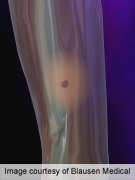
Visual images such as those of benign and cancerous skin lesions increase awareness and accuracy of skin self-examination, according to a review published in the July issue of the Journal of the American Academy of Dermatology.
(HealthDay)—Visual images such as those of benign and cancerous skin lesions increase awareness and accuracy of skin self-examination, according to a review published in the July issue of the Journal of the American Academy of Dermatology.
Jennifer E. McWhirter and Laurie Hoffman-Goetz, Ph.D., M.P.H., from the University of Waterloo in Canada, identified and reviewed outcomes from 25 published studies that used visual images to promote skin self-examination for early detection of skin cancer.
The researchers found that visual images increased knowledge and self-efficacy related to skin self-examination and increased the frequency and accuracy of skin self-examination and melanoma detection. The most effective images were mole-mapping diagrams, baseline photographs of patients' own moles and skin surfaces, benign and cancerous lesions, dermoscopy, and lesions changing over time. Text descriptors alone were found to be ineffective.
"Evidence from this systematic review suggests an important role for visual images in patient education related to informed self-monitoring for skin lesions," McWhirter and Hoffman-Goetz conclude. "Patients should have access to images for viewing at any point in time, and to large quantities of exemplars, to augment visual memory and pattern-based recognition, respectively."
More information:
Abstract
Full Text (subscription or payment may be required)
Journal information: Journal of the American Academy of Dermatology
Health News Copyright © 2013 HealthDay. All rights reserved.


