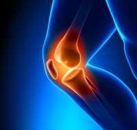
New research from the University could help improve the success of knee replacement surgery and prevent costly revisions to existing implants.
A new study from researchers at the University has identified a more accurate computerised method that will improve the success of knee replacements, and prevent costly, often unnecessary revisions to existing surgical implants.
Modern knee replacement is a highly successful operation, relieving the pain and disability of knee osteoarthritis. Osteoarthritis is the degeneration of joint cartilage and underlying bone, particularly affecting the knee, hip and thumb joints and is the world's most common musculo-skeletal disease.
It usually affects middle-aged men and women from 40 years, causing pain and stiffness, usually worsening with time. By 2020, the World Health Organisation predicts osteoarthritis will be the world's fourth leading cause of disability, affecting double the number of patients who have cardiac disease. Studies suggest that almost one in two people will get symptomatic osteoarthritis of the knee during their lifetime.
Every year 90,000 knee replacements are performed in the UK, a figure likely to rise by more than 600 per cent by 2030 due to an ageing population, increased obesity and the fact that knee replacement patients are getting younger. This in turn, increases the number of patients requiring a second knee replacement or 'revision' in their lifetime.
But the surgery has limitations however; along with population changes, the move to patient-led decision making and the threat of antibiotic resistance present significant challenges for current and future healthcare providers. Revisions to knee replacements can cost up to four times the amount of the original surgery, and bring increased risk of infection and failure.
Radiolucency is a region surrounding a hip or knee replacement which is dark on an X-ray and if it worsens with time can indicate implant loosening. In severe cases, the implant may need to be revised. In current clinical practice radiolucency is assessed visually by orthopaedic surgeons; this is therefore qualitative and agreement between clinicians is poor.
In this study, researchers developed a semi-automated computer program which could be run by an unskilled user and which provided an independent, more reliable radiolucency score than the present subjective surgeons' assessments. If used clinically, this tool would improve diagnosis and limit potentially unnecessary surgical revisions.
The study, published in Interface, the Journal of the Royal Society, analysed six surgeons' assessment of radiolucency in 38 unicompartmental knee replacement radiographs. The results were then compared with assessments made by a semi-automated imaging algorithm. While large variation was found between the surgeon results, with total agreement in less than 10 per cent of zones, the automated programme had total agreement in 81.6 per cent of zones – demonstrating a more accurate and reliable means of diagnosing radiolucency.
"Surgeons are given limited guidance on how to define radiolucency and use different assessment criteria which explains the wide and concerning variation found in the surgical assessments in this study," commented Richie Gill, Professor of Healthcare Engineering within our Department of Mechanical Engineering.
"Using a digital computerized tool that accurately identifies patients with progressive pathological radiolucency – severely loosening knee replacements – would ensure correct surgical procedures are applied, improving patient outcomes and saving money spent on operations which may not ultimately be successful."
Mr Senthil Kumar Ganesan, orthopaedic surgeon at the Royal United Hospital in Bath said: "Total knee replacements are on the increase mainly due to the increasing demand from the patients as well as younger patients under the age of 60, undergoing knee replacements.
"As the younger patients are more physically demanding and also live longer, the average expectancy of the knee replacements is 10-15 years. These patients will require revision surgery in the 70s and further surgery in their 80s. This makes the operation very technically challenging and also requires more hardware to do the operation. The use of early and accurate diagnostic tools in the diagnosis of early loosening will be of immense help in the knee replacement surgery. This will not only facilitate early treatment it will also help to prevent late complications of neglected loosening."
More information: E. C. Pegg, B. J. L. Kendrick, H. G. Pandit, H. S. Gill, and D. W. Murray. "A semi-automated measurement technique for the assessment of radiolucency." J. R. Soc. Interface July 6, 2014 11 96 20140303; DOI: 10.1098/rsif.2014.0303 1742-5662
Journal information: Journal of the Royal Society Interface
Provided by University of Bath


