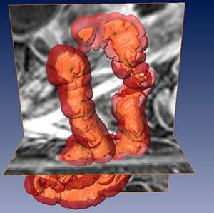
Credit: Delft University of Technology
Researchers Robiël Naziroglu and Frans Vos have developed a method for improving the assessment of MRI images in cases of Crohn's disease. In time, this may well enable more targeted monitoring and treatment of this chronic intestinal inflammation. Naziroglu will take his doctorate on this subject at TU Delft on Monday 21 November.
Better targeting
Medical imaging techniques have become essential for the assessment of all kinds of disorders. The researcher Robiël Naziroglu studied how imaging techniques could be used to improve the assessment of Crohn's disease, in principle making it possible to treat the illness in a more targeted way. TU Delft has been active for some years in the field of improved imaging techniques for intestinal research, among other things, such as mapping the presence of polyps.
700,000 patients in Europe
Crohn's disease is a chronic inflammatory disease of the gastrointestinal tract that occurs relatively often in the Western world. In Europe alone, around 700,000 people suffer from the condition. 'The disease is characterised by regular alternations between active and less active phases,' says Naziroglu. 'This means that it is crucial to determine the activity level as well as the presence of the disease. This disease activity can be divided into different score levels, and every score level has a different treatment plan. This so-called grading of the Crohn's disease activity is therefore important for monitoring the progression of the disease and for determining the treatment strategy and whether it is working.'
A better scoring system
'Ideally, such a score for disease activity would be objective, reproducible, quantitative, non-invasive and complete. Unfortunately, however, less than perfect scoring systems are still being used at present, based on MRI and endoscopy, among other things. The aim of the VIGOR++ project, of which my research formed a part, was to develop a better scoring system by looking at both manually and automatically obtained MRI measurements.'
Complex
This proved a challenge. 'That's because the intestines are relatively complex, including in a geometric sense. The segmentation (division) of the intestinal wall is a crucial step in being able to identify the disease automatically from Crohn's activity on MRI images, but it's therefore tricky. So I developed an algorithm that is able to handle the complexity of the intestines well, by using prior knowledge of the content of the intestines and of knowledge of the surrounding anatomy.'
Testing
The method was developed using images from 27 patients with Crohn's disease and subsequently tested on 120 patients. 'Our semi-automatic method allows us to obtain very reproducible outlines and thickness measurements of actively diseased parts of the intestinal wall. With the VIGOR++ system, a method was also developed to score and validate disease activity using endoscopy. As well as this, the system was compared with other state-of-the-art MRI scoring systems and was shown to be performing well: the reproducibility proved to be significantly higher.'
Hospitals
For his research, Naziroglu worked closely with doctors, including at the Academic Medical Center (AMC) in Amsterdam. It remains unclear as to the extent to which, and how often, his method will be used in the medical world. 'The method will certainly have to be tested further and developed before it can be used in practice on a large scale, and adjustments will need to be made, but I think that this new method is very promising.'
Provided by Delft University of Technology


