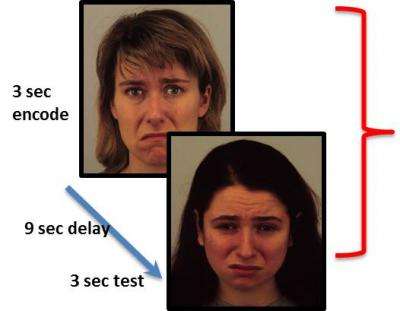
Over several trials, participants were required to attend to either the identity (non-emotional feature) or the emotion of a face, remember it during a 9-second delay, and match the feature to a subsequent face. Neural activity in the visual cortex elicited by the emotion trials predicted depressed patients' subsequent responsiveness to scopolamine treatment. Credit: Maura Furey, Ph.D., NIMH Experimental Therapeutics and Pathophysiology Branch
(HealthDay)—The neural response in the visual cortex while processing emotional information can predict which patients with major depressive disorder will respond to scopolamine, according to a study published online Jan. 30 in JAMA Psychiatry.
To determine whether neural activity when processing emotional information could predict the antidepressant response to scopolamine, Maura L. Furey, Ph.D., from the National Institutes of Health in Bethesda, Md., and colleagues performed functional magnetic resonance imaging on 15 adults with major depressive disorder and 21 healthy adults. The images were taken while they performed face-identity and face-emotion working memory tasks after the administration of placebo or scopolamine.
NIMH's Dr. Maura Furey talks about how a functional brain imaging measure may help predict a patient's response to a rapid-acting experimental antidepressant. Credit: National Institute of Mental Health
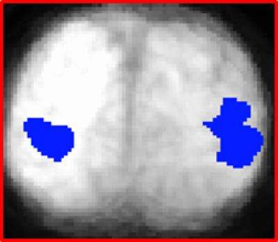
The researchers found that the baseline blood oxygen level-dependent response in the bilateral middle occipital cortex during the processing of emotional information correlated with the magnitude of the response to scopolamine. The change in neural activity in overlapping areas in the middle occipital cortex during stimulus-processing components while performing the emotion task after receiving scopolamine also correlated with treatment response. During baseline, healthy controls exhibited higher activity in the same visual regions compared with patients with major depressive disorder.
The level of increased activity in left and right visual cortex (blue), while attending to emotional faces in a working memory task, predicted depressed patients' responsiveness to the experimental antidepressant scopolamine. View from the back of the brain shows fMRI data superimposed on anatomical MRI scan data. Credit: Maura Furey, Ph.D., NIMH Experimental Therapeutics and Pathophysiology Branch
"These results implicate cholinergic and visual processing dysfunction in the pathophysiology of major depressive disorder and suggest that neural response in the visual cortex, selectively to emotional stimuli, may provide a useful biomarker for identifying patients who will respond favorably to scopolamine," Furey and colleagues conclude.
Several authors have patent applications for the use of scopolamine and ketamine for the treatment of depression.
More information:
Abstract
Full Text (subscription or payment may be required)
Journal information: JAMA Psychiatry
Health News Copyright © 2013 HealthDay. All rights reserved.


