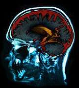
(HealthDay)—Patients with posttraumatic stress disorder (PTSD) exhibit alterations in the topological architecture of the brain, according to a study published in the September issue of Radiology.
Du Lei, Ph.D., from the West China Hospital of Sichuan University in Chengdu, and colleagues used magnetic resonance imaging (MRI) and graph theory approaches to examine the topological organization of the functional connectome of patients with PTSD. Seventy-six patients with PTSD caused by an earthquake and 76 matched control subjects who experienced the same disaster underwent resting-state functional MRI.
The researchers found that patients with PTSD exhibited abnormalities in global properties, including a significant decrease in path length (P = 0.0002), and increases in the clustering coefficient, global efficiency, and local efficiency (P = 0.0014, 0.0002, and 0.0004, respectively), compared with control subjects. Increased centrality in nodes mainly involved in the default mode network (DMN) and the salience network (SN), including the posterior cingulate gyrus, precuneus, insula, putamen, pallidum, and temporal regions, were exhibited locally by patients with PTSD.
"These results suggest that individuals with PTSD exhibit a shift toward 'small-worldization' (in which the network transforms from a random or regular network to a small-world network) rather than toward randomization; furthermore, the disequilibrium between the DMN and the SN might be associated with the pathophysiology of PTSD," the authors write.
Copyright © 2015 HealthDay. All rights reserved.


