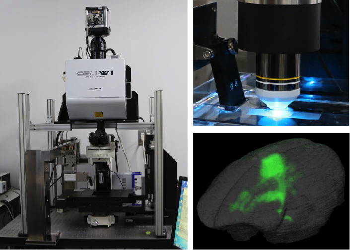
AST setup and whole-brain imaging in mouse and marmoset. Credit: Osaka University
Researchers at Osaka University develop a high-speed serial-sectioning imaging system that captures high-resolution images of a whole mouse brain and furthers our understanding of brain diseases in rodents and primates
To fully understand brain function and dysfunction, it is important to be able to visualize changes in anatomy and activity in the whole brain. High-resolution brain imaging that can distinguish individual cells and quantitative comparison of acquired data are essential to show how the brain is affected by disease.
However, current attempts to image a whole mouse brain at a resolution high enough to gain detailed information take up to one week. While these approaches have revealed important insights into brain function, it is not possible to image and analyze multiple brains with these technologies. Comparing multiple brains is essential to understand neurobiological function and dysfunction in brain disorders.
Osaka University-led researchers have developed block-face serial microscopy tomography (FAST)—an imaging system that can image a whole mouse brain at high spatial resolution in less than two-and-a-half hours. "FAST consists of a spinning disk confocal microscope with built in microslicer and a method for processing image data," explains first author Kaoru Seiriki. "With our 3-D reconstruction technique, whole brains can be visualized at a resolution high enough to resolve individual cells and their subcellular structures."
By combining their FAST technique with specific staining procedures, Seiriki and colleagues were able to visualize subcellular nuclei, vascular structures, mature oligodendrocytes, myelin sheaths, interneurons, and projecting neurons throughout the whole brain. These imaging tools provide a systemic approach to investigating the pathophysiological mechanisms of different brain diseases.
FAST is a very quick imaging technique, therefore it can potentially be used to image non-human primate brains. "We successfully visualized a long-range neuronal projection at a subcellular resolution in the whole brain of an adult marmoset," says leading researcher Hitoshi Hashimoto. "This shows how FAST can further our understanding of brain anatomy in rodents and primates."
The Osaka University research team has also successfully imaged postmortem human brains using their FAST system. "We expect that this approach will identify fine morphological abnormalities in diseased human brains that were previously unknown," says Hashimoto. These insights may help to advance the development of effective treatments.
This new technique offers a way to compare multiple brains at the level of individual cells and their subcellular structures. With this approach, new insights will be gained into the pathological mechanisms of different brain diseases. Furthermore, the applicability of this offers a translational approach to researching non-human animal models and human diseases.
More information: Kaoru Seiriki et al. High-Speed and Scalable Whole-Brain Imaging in Rodents and Primates, Neuron (2017). DOI: 10.1016/j.neuron.2017.05.017
Journal information: Neuron
Provided by Osaka University


