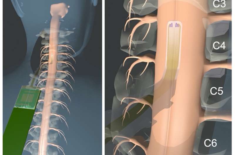
Diagram illustrating the placement of the array on the participant’s spinal cord. Credit: S.M. Russman, et al., Science Translational Medicine (2022)
A combined team of researchers from the University of California and Oregon Health and Science University, has developed a microelectrode array to precisely monitor electrophysiology during spinal surgery. They have published their work in the journal Science Translational Medicine.
Spinal surgery is a delicate operation. Surgeons not only have to fix what is wrong, but must do so without damaging nerves, which can lead to impairments in patients. One area of particular concern is tumor removal, but even just conducting biopsies is risky. Spinal surgeons currently use intraoperative neuromonitoring devices that monitor electrical activity in the nervous system. With such devices, electrodes are placed on the back muscles and the scalp. Unfortunately, they do not provide precise information, which is what surgeons need. In many instances, they serve only to alert the surgeon to damage that has already occurred. In this new effort, the researchers have attempted to improve on the system by creating a device that allows for more precise electrophysiology monitoring during surgery.
The new device consists of an array of electrodes that the surgeon places directly onto the spinal column near the main surgery site. The electrodes detect electrophysical responses from the spinal nerves and transmit them to a computer that coverts them to a submillimeter-precision 2D map, which the surgeon can consult in real time as they carry out a surgical procedure.
The time evolution of the response, phase gradients, and streamlines during SSEP monitoring. Credit: S.M. Russman, et al., Science Translational Medicine (2022)
The researchers tested their new device during six actual procedures on volunteers. They found that their device provided higher sensitivity than current devices and outperformed them in assisting the surgeon during spinal surgeries.
More testing is required before the device can be submitted for approval, but the researchers are confident their system will provide more accurate information to spinal surgeons and thus prevent neural damage in future patients. They also suggest that their device could be used to monitor the progress of healing in people with damaged spinal columns.
More information: Samantha M. Russman et al, Constructing 2D maps of human spinal cord activity and isolating the functional midline with high-density microelectrode arrays, Science Translational Medicine (2022). DOI: 10.1126/scitranslmed.abq4744
Journal information: Science Translational Medicine
© 2022 Science X Network


