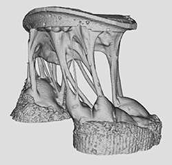
Example of the high level of detail of the mitral valve structures obtainable by 7T MRI imaging. Ventricular views displayed, showing the papillary muscles and chordae tendineae. The valve is submerged in fluid and in a zero-pressure natural state. Credit: University of Arkansas
On Aug. 30, a team led by associate professor of biomedical engineering Morten Jensen published an article in the scientific journal PLOS ONE titled "High resolution imaging of the mitral valve in the natural state with 7 Tesla MRI." The article discusses a study in which Jensen and colleagues sought to develop a method and apparatus for obtaining high-resolution 3D MRI images of porcine mitral valves. The systematic approach taken by the authors produced 3D datasets of high quality which, when combined with physiologically accurate positioning by the apparatus, can serve as an important input for validated computational models.
"This is the first time that anyone has been able to produce 3D image datasets with MRI of this high quality, resolution and S/N ratio of a heart valve in its natural, relaxed state surrounded by fluid," said Jensen. "It is an important step in itself to understand the valve structures better, but especially important for computational biomechanical modeling with force boundary conditions from the exact same valve."
"We are excited at the partnerships being developed by Dr. Jensen with the FDA and other SEC institutions. This work is a great example of how perspectives from computational modeling, biomechanics and imaging modalities come together to address critical questions in the functioning of the cardiovascular system," added Raj Rao, professor and head of the department of biomedical engineering.
More information: Sam E. Stephens et al. High resolution imaging of the mitral valve in the natural state with 7 Tesla MRI, PLOS ONE (2017). DOI: 10.1371/journal.pone.0184042
Journal information: PLoS ONE
Provided by University of Arkansas


