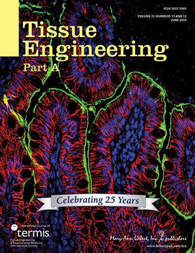
Credit: (c) 2019 Mary Ann Liebert, Inc., publishers
The loss of complete segments of the esophagus often results from treatments for esophageal cancer or congenital abnormalities, and current methods to re-establish continuity are inadequate. Now, working with a rat model, researchers have developed a promising reconstruction method based on the use of 3-D-printed esophageal grafts. Their work is published in Tissue Engineering.
Eun-Jae Chung, MD, Ph.D., Seoul National University Hospital, Korea, Jung-Woog Shin, Ph.D., Inje University, Korea, and colleagues present their research in an article titled "Tissue-Engineered Esophagus via Bioreactor Cultivation for Circumferential Esophageal Reconstruction". The authors created a two-layered tubular scaffold with an electrospun nanofiber inner layer and 3-D-printed strands in the outer layer. After seeding human mesenchymal stem cells on the inner layer, constructs were cultured in a bioreactor, and a new surgical technique was used for implantation, including the placement of a thyroid gland flap over the scaffold. Efficacy was compared with omentum-cultured scaffolding technology, and successful implantation and esophageal reconstruction were achieved based on several metrics.
"Dr. Chung and colleagues from Korea present an exciting approach for esophageal repair using a combined 3-D printing and bioreactor cultivation strategy," says Tissue Engineering Co-Editor-in-Chief John P. Fisher, Ph.D., Fischell Family Distinguished Professor & Department Chair, and Director of the NIH Center for Engineering Complex Tissues at the University of Maryland. "Critically, their work shows integration of the engineered esophageal tissue with host tissue, indicating a clinically viable strategy for circumferential esophageal reconstruction."
More information: In Gul Kim et al. Tissue-Engineered Esophagus via Bioreactor Cultivation for Circumferential Esophageal Reconstruction, Tissue Engineering Part A (2019). DOI: 10.1089/ten.tea.2018.0277
Provided by Mary Ann Liebert, Inc


