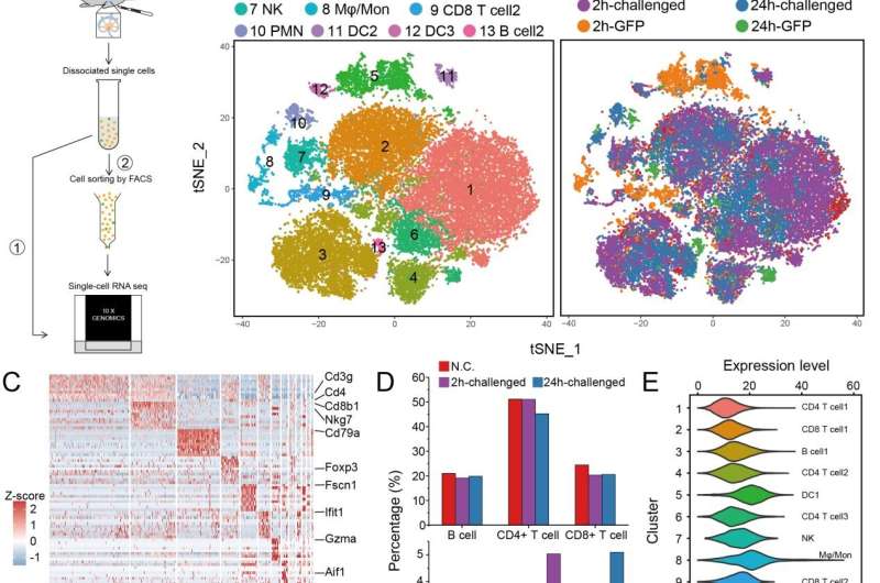
Single-cell RNA sequencing was performed in the lymph nodes of mice at 2h and 24h post infection with Y. pestis, which showed that inflammation related pathway genes were significantly up-regulated in the early stage of infection. Compared with T cells and B cells, innate immune cells tended to interact with Y. pestis during the early stage of infection. Credit: Science China Press
This study was led by Dr. Ruifu Yang, Dr. Zongmin Du (State Key Laboratory of Pathogen and Biosecurity, Beijing Institute of Microbiology and Epidemiology) and Dr. Fan Bai (Biomedical Pioneering Innovation Center, School of Life Sciences, Peking University).
Yersinia pestis (Y. pestis), the causative agent of plague, has caused millions of deaths in three worldwide pandemics in history. Plague is predominantly a flea-borne zoonotic disease. While only sporadic human plague outbreaks occur around the world each year, the threat of plague to humans and society should not be underestimated.
Three subtypes of the disease are commonly encountered in the clinic, namely bubonic, septicemic, and pneumonic plagues. Bubonic plague is the major form and is characterized by enlarged purulent abscesses called "buboes", which are caused by the massive replication of Y. pestis after its rapid migration into the dLNs post infection. Relevant research on paralysis of the innate immune responses by Y. pestis during the early stages of infection is limited, which has hindered our understanding of Y. pestis pathogenesis.
The researchers challenged mice with fluorescent labeled Y. pestis by subcutaneous injection into the groins of mice to simulate bubonic plague development. On this basis, they took mouse inguinal lymph nodes at the 2nd hour and the 24th hour post infection (hpi) and analyzed the changes in the composition and status of immune cells in the lymph nodes by flow cytometry and single-cell transcriptome sequencing.
Compared with the control group, genes of the inflammation related pathway were significantly up-regulated in the lymph node cells of mice infected with Y. pestis during the early stage of infection. The researchers calculated the infection preference index by the proportion of specific fluorescent labeled cells and found that Y. pestis tend to interact with the resident innate immune cells including dendritic cells, monocytes/macrophages and neutrophils, rather than adaptive immune cells.
-
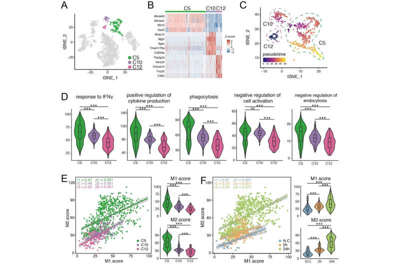
Monocytes/macrophages infected with Y. pestis exhibited an M1-M2 coupled activation state rather than the classic M1/M2 polarization modes. Credit: Science China Press
-
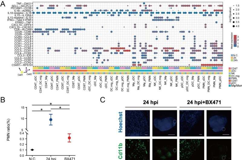
Neutrophils that play vital roles in eliminating of Y. pestis were enriched in dLNs at 24 hpi, which was the results of the interactions between chemokines secreted by other cells in dLNs and chemokine receptor CCR1. Credit: Science China Press
Subsequently, the researchers further sorted the resident innate immune cells and performed the scRNA-seq. It was found that monocytes/macrophages showed M1 and M2 coupling activation in response to Y. pestis infection, which could be the comprehensive results of the activation of immune response mediated by host pattern recognition receptors and the inhibition effects of various virulence factors of Y. pestis.
In addition, the team also used the cell interaction analysis tool based on CellPhoneDB to reveal the ligand receptor interaction of different cell types at different infection time points. It was found that the chemokine receptor CCR1 of neutrophils may be involved in the recruitment of neutrophils to lymph nodes post infection, and the CCR1 function inhibition experiments were performed to verify this conclusion.
Taken together, this study demonstrated the critical cell types involved and critical immune events occurred in the initial interactions between Y. pestis and the host at the single-cell transcription level, which will be valuable for understanding the development of bubonic plague and host immune response to Y. pestis at the early stage.
The research was published in Science China Life Sciences.
More information: Yifan Zhao et al, Single-cell transcriptomics of immune cells in lymph nodes reveals their composition and alterations in functional dynamics during the early stages of bubonic plague, Science China Life Sciences (2022). DOI: 10.1007/s11427-021-2119-5
Journal information: Science China Life Sciences
Provided by Science China Press


