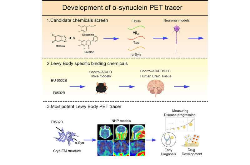
The discovery of F0502B represents a promising lead compound for imaging α-synuclein inclusions in synucleinopathies. Credit: Ye Keqiang
Brain imaging scans are powerful tools for diagnosing Parkinson's disease (PD) and ruling out other motor disorders. In PD, the presence of α-synuclein (α-Syn) in Lewy bodies and Lewy neurites in the brain, particularly the substantia nigra, distinguishes it from other parkinsonisms. Unfortunately, there is currently no effective α-Syn PET tracer available.
Now, a research team led by Prof. Ye Keqiang from the Shenzhen Institute of Advanced Technology (SIAT) of the Chinese Academy of Sciences has discovered a promising compound called F0502B for imaging α-Syn and aiding in the diagnosis of synucleinopathies. The study was published in Cell on July 7.
The researchers found that in monkey PD models, [18F]-labeled F0502B specifically bound to α-Syn fibrils with high affinity, distinguishing them from Aβ and Tau fibrils.
PET imaging in monkey models showed that [18F]-F0502B specifically detected α-Syn aggregates. The levels of aggregated α-Syn may increase over time, leading to elevated PET-specific binding signals.
F0502B can act as neuroimaging radiotracer due to its certain chemical and pharmacological properties, including high permeability through the blood-brain barrier, rapid clearance from normal brain tissue and blood, and high-affinity binding and selectivity for the target.
"Our findings demonstrate that F0502B acts as a specific PET tracer by selectively binding to aggregated α-Syn," said Prof. Ye, corresponding author of the study. "This could enhance our understanding of disease progression and potentially facilitate monitoring therapeutic efficacy in clinical trials."
More information: Jie Xiang et al, Development of an α-synuclein positron emission tomography tracer for imaging synucleinopathies, Cell (2023). DOI: 10.1016/j.cell.2023.06.004
Journal information: Cell
Provided by Chinese Academy of Sciences


