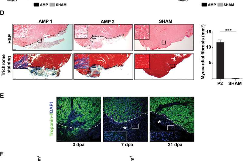
Scar and new myocardial tissue assessment following surgical amputation of the left ventricular apex in P2 mice revealed a lack of myocardial regeneration. (A) Survival of P2 mice that underwent amputation (AMP; n = 91) or not (SHAM; n = 51) of a small portion of the left ventricular apex. Animal survival was determined immediately (0 h), after 3 hours (3 h), and the day after surgery (24 h). Data are means ± SEM. (B) Stereomicroscopic pictures of AMP and SHAM hearts at 21 dpa demonstrated the presence of extensive white patches on the external ventricular wall 21 days following surgery (black arrowheads). Scale bar, 1 mm. (C) Heart weight of P2 AMP (n = 3) and SHAM (n = 3) mice was determined at different time points following surgery. (D) Representative H&E- and trichrome-stained sections of P2 hearts at 21 dpa. Cardiac tissue loss is evident by the misalignment of tissue fibers (H&E inset) and by the presence of intense blue staining (trichrome inset). Dashed line marks the lesion border. Scale bar, 200 μm. Right: Quantification analysis of the scar area in P2 AMP versus SHAM at 21 dpa. ***P < 0.001. (E) Paraffin-embedded resected P2 hearts were sectioned and immunostained for troponin-I and DAPI. Representative images at various time points during the post-AMP process demonstrate an absence of newly formed myocardial tissue. An asterisk indicates the healing area, whereas the dashed line marks the lesion border. Scale bar, 25 μm. (F) Number of proliferating CMs (DAPI+/MF-20+/BrdU+ cells) per 0.1 mm2 counted in paraffin-embedded SHAM and AMP heart sections from BrdU multiple pulse-chase experiments at the indicated dpa. Data are means ± SEM; n = 3. Only fields completely filled with cells were included in the analysis. ns, not significant. Credit: Science Advances (2018). DOI: 10.1126/sciadv.aao5553
A team of researchers from several institutions in Spain has found that heart muscle regeneration in neonatal mice only occurs during the first day after birth. In their paper published on the open access site Science Advances, the group explains their study of regeneration of heart muscle in newborn mice and what they found.
Back in 2011, a team of researchers claimed to have seen heart muscle regeneration in newborn mice. They reported that after clipping off a piece from a tiny heart, the muscle would regenerate, a form of self-repair. The report caused a stir, because no other researchers had ever seen such heart muscle rejuvenation in mammals. It also caused some controversy, as some researchers were unable to replicate the experiments while others succeeded. In this new effort, the researchers sought not only to replicate those results, but to better understand the variable efforts at replication.
The experiments by the team were straightforward and simple. They opened up several healthy, newly born mice, and cut off a piece of the heart muscle in a non-lethal procedure. The procedure was performed on mice that were one through four days old, and nine days old. They then closed up the mice and allowed them to heal for three weeks. At that point, they opened them up again to see if the heart muscle had regenerated. The researchers report that their findings were not so simple. They report that the mice who had lost heart muscle on day one showed minimal signs of scarring. All of the others had significant scarring where the heart muscle had been snipped. The researchers suggest that the heart muscle on the day one mice regenerated, but as yet, they cannot prove it. All they can say for sure is that there was little scarring and that the hearts of those mice were the same size and weight as unaltered control mice.
To uncover the reason for the differences between day one mice and all the others, the researchers looked at factors that involve gene expression for muscle stiffness in developing mice. They found that it was significantly higher in newborn mice after the first day. They then gave test mice a drug that prevented such stiffness from occurring and reran the initial experiments. Doing so, they report, resulted in all of the mice showing very little scarring.
More information: Mario Notari et al. The local microenvironment limits the regenerative potential of the mouse neonatal heart, Science Advances (2018). DOI: 10.1126/sciadv.aao5553
Abstract
Neonatal mice have been shown to regenerate their hearts during a transient window of time of approximately 1 week after birth. However, experimental evidence for this phenomenon is not undisputed, because several laboratories have been unable to detect neonatal heart regeneration. We first confirmed that 1-day-old neonatal mice are indeed able to mount a robust regenerative response after heart amputation. We then found that this regenerative ability sharply declines within 48 hours, with hearts of 2-day-old mice responding to amputation with fibrosis, rather than regeneration. By comparing the global transcriptomes of 1- and 2-day-old mouse hearts, we found that most differentially expressed transcripts encode extracellular matrix components and structural constituents of the cytoskeleton. These results suggest that the stiffness of the local microenvironment, rather than cardiac cell-autonomous mechanisms, crucially determines the ability or inability of the heart to regenerate. Testing this hypothesis by pharmacologically decreasing the stiffness of the extracellular matrix in 3-day-old mice, we found that decreased matrix stiffness rescued the ability of mice to regenerate heart tissue after apical resection. Together, our results identify an unexpectedly restricted time window of regenerative competence in the mouse neonatal heart and open new avenues for promoting cardiac regeneration by local modification of the extracellular matrix stiffness.
Journal information: Science Advances
© 2018 Medical Xpress


