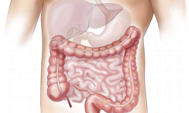
Credit: CC0 Public Domain
Colonic diverticulosis is a prevalent condition among older adults, marked by the presence of thin-walled pockets in the colon wall that can become inflamed and infected; they can then hemorrhage or rupture. A new study, published in the journal Gut now suggests that tissue remodeling is a primary mechanism for diverticula formation.
The study, led by corresponding author Anne Peery, MD, associate professor of medicine in the Division of Gastroenterology and Hepatology at the UNC School of Medicine, aimed at identifying the genetic and cellular determinants underlying colonic diverticulosis and the relationship with other gastrointestinal disorders.
Researchers conducted DNA and RNA sequencing on colonic tissue from 404 patients. Results showed 38 genes with differential expression and 17 with varied transcript usage linked to diverticulosis. Additionally, diverticulosis severity was positively correlated with genetic predisposition to diverticulitis.
Peery and colleagues linked the formation of diverticula to stromal and epithelial cells in the colon, particularly endothelial cells, myofibroblasts, fibroblasts, goblet, tuft, enterocytes, neurons, and glia. The researchers also analyzed genes to find five—CCN3, CRISPLD2, ENTPD7, PHGR1 and TNFSF13—that showed potential causal effects on diverticulosis. Interestingly, the researchers confirmed that the gene ENTPD7 was over-expressed in diverticulosis cases.
In addition, the study showed that individuals with an increased genetic proclivity to diverticulitis show larger numbers of diverticula upon colonoscopy.
More information: Jungkyun Seo et al, Genetic and transcriptomic landscape of colonic diverticulosis, Gut (2024). DOI: 10.1136/gutjnl-2023-331267


