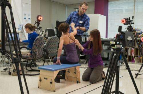
Marking the spot: Collaboration aims to develop clinically useful tool to shed light on birth injury

University of Delaware researcher Jim Richards has successfully used motion analysis technology to allow elite figure skaters to explore "what-if" scenarios about their jumping technique. Now he hopes that he and his research team can use a similar approach to guide clinicians in treating children with a birth injury called brachial plexus birth palsy (BPBP).
BPBP, which occurs in about four out of every 1,000 births, affects nerve roots in the cervical spine, impacting muscle function in the shoulder and the arm. Most children recover on their own, but about 30 percent are left with lifelong deficits in arm function that require therapy or surgery. The most severe brachial plexus injuries can cause complete paralysis of the arm.
But the answer to a key question has eluded researchers trying to understand exactly what is going on in the musculoskeletal systems of children with BPBP: Where is the shoulder blade at any given moment, and what is it doing? This information would provide valuable insight into a child's specific defects and enable treatments to be tailored to individual patients, as the location and extent of damage to the nerves and muscles vary from one person to another.
Richards, who is Distinguished Professor in UD's Department of Kinesiology and Applied Physiology, explains that the movements of the scapula, commonly known as the shoulder blade, are incredibly difficult to measure.
"Our motion capture cameras provide us with reasonable data for the lower extremities," Richards says, "but the same approach applied to the upper body fails to tell us much about the movement of the scapula."
He and his team of doctoral students in UD's BIOMS (Biomechanics and Movement Science) program have taken a systematic approach to filling this gap. If they're successful, it may someday be possible for surgeons to use the UD simulation to explore what will happen if they move a tendon from one point to another in an individual patient.
Feeling their way
The team is working with clinicians at Shriners Hospital for Children in Philadelphia on the first stage of the project. Two of Richards' students, Kristen Thomas and Stephanie Russo, have collected data on 65 children with BPBP using a motion analysis system.
"We're fairly confident that we can get accurate scapular measurements under static conditions," Thomas explains. "The question then is, 'If we put the kids in enough static positions, can we draw conclusions about what happens when they're moving?'"
"There are 11 specific positions that have clinical relevance," she continues. "The problem is that right now we're identifying these positions through palpation, or feel, and the accuracy of this approach has not been established in living patients. So our plan is to use static fluoroscopic imaging data to build a 3D model for comparison with the palpation method. If the model validates the palpation measures, then we can move forward with testing a larger pool of subjects without the need for expensive imaging equipment."
Doing the math
Once the researchers have determined whether the positional measurements are repeatable, they can develop a set of equations to tell them how the scapula moves from one position to another. Results from the equations will be compared with data collected using 3D motion fluoroscopy, an imaging technique that produces a video X-ray.
"If it all works," Richards says, "we'll be able to go into a clinical setting like Shriners, drop 11 markers onto a patient to find out what's happening, and then do the same after surgery to see what the effects are."
Seeing the big picture
Tyler Richardson, another graduate student in Richards' group, is adapting a freely available program called OpenSim for use on the project.
"With motion data input from a patient, the program will use mathematical optimization to find all of the possible muscle combinations that could produce that motion," he says. "We can gain lots of information about the muscle function, both pre- and post-surgically, of an individual this way."
Ultimately, Richardson envisions clinicians being able to explore what-if scenarios that would enable them to determine how a specific surgical technique will affect a specific patient. And eventually the technique could be broadened beyond BPBP to other injuries.
The academic researcher has high praise for his clinical partners on the project as well as his for his team of talented students.
"Shriners is the go-to place for families who have a child with this injury," he says. "We're very fortunate to be working with these experts, and none of this would be possible without my grad students."
Russo, who is simultaneously working on a Ph.D. at UD and an M.D. at Drexel University, is co-advised by two physicians at Shriners, Scott Kozin and Dan Zlotolow.
"They have been fabulous to work with," she says. "It's wonderful to see the collaboration between the science side through UD's BIOMS program and the clinical side through the physicians at the hospital. We're all working together to make treatment more effective for these kids."


