Lab findings put patients on sound footing

Imagine being able to look inside the body in motion, and see not only the biomechanics of bones and joints, but also their interaction with the outside world.
Western researchers are working on perfecting just that.
Not too long ago, Tom Jenkyn examined how the body moved by taking multiple-angle, optical motion shots with track markers on the skin.
Working in Western's Wolf Orthopaedic Biomechanics Laboratory, at the Fowler Kennedy Sports Medicine Clinic, Jenkyn, the lab's co-director, used a dozen cameras to capture a patient's movement to understand conditions ranging from joint problems to sport injuries.
"Since 2002, at Fowler, we've used optical motion cameras to capture (movement) with track markers on the skin. That kind of technology was developed for people like me, to see how people move," said Jenkyn, who is cross appointed with the faculties of of Engineering and Health Sciences.
"It's the kind of system you see in computer graphics or video games. One drawback is you're sticking markers to the skin, but you really want to know what bones are doing under the skin. We need to look at bones directly," he said.
Seeing actual bones and joints in motion could help researchers and health-care providers, such as physiotherapists, better understand injuries, how to foresee them, how to prevent them and how well patients respond to therapeutic treatment.
To accomplish this, Jenkyn has found a new way to examine the body's biomechanics beneath the surface. His method of looking at bones and joints in motion, beneath the skin, is the first of its kind in Canada.
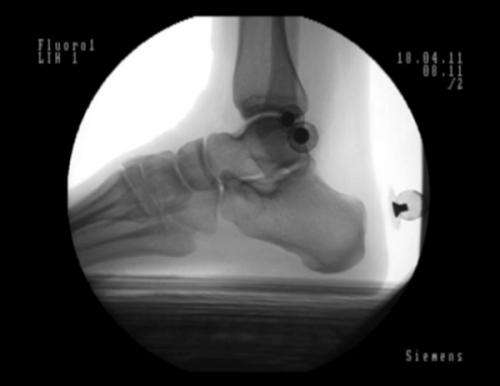
In order to look at bones directly, Jenkyn and his team, in the relatively new Wolf Orthopaedic Quantitative Imaging Laboratory (WOQIL), thought of paring three-dimensional images with X-rays, in order to get a better understanding of what's going on beneath the surface.
"We settled on X-ray fluoroscopy, a moving X-ray, the kind surgeons use when putting a catheter into a heart, to see what's inside. When you look at a joint or bone from two different angles, you can triangulate its position and orientation and this gives you the info that you need," said Jenkyn, WOQIL director.
"It works great, but your view volume is very small – a 9-inch window into the body. So, we do (fluoroscopy) at the same time as the optical shots with markers. It gives us overall motion, so we stick the X-ray on the joint we're interested in and it gives us data with a very high accuracy, of 0.05mm," he continued.
"If you're looking for things like why some people get osteoarthritis and some don't, if you're looking for a biomechanical cause, that's the kind of accuracy we need."
WOQIL has six digital cameras, which take pictures of the body's movement on the outside, and two three-dimensional fluoroscopic radiostereometric analysis machines that show what's happening inside with the aid of an X-ray.
Jenkyn's work could help improve understanding and treatment of a wide array of sports injuries and joint problems, even one's posture and how the spine changes when you slouch.
"We have a shoulder study right now going on, looking at how the shoulder moves abnormally in rotator cuff tears, how your body compensates," he noted, adding data collected can help them examine the healing process and how the patient is responding to physiotherapy.
"Surgery may not have gone as well but physio can save you. But 'physios' are working a bit blind; they're not sure if the bone is going back, so we can give them that info."
The lab has recently looked at the foot and how orthotics work for flat feet.
"We had questions about what the orthotics were doing, why they don't work for everybody. The nice thing about the fluoro is, we can see bones in foot, the orthotic, so we've been experimenting with looking at how orthotics are working and designing new ones based on that," he explained.
That's just one real-world application; the lab can look at materials outside the body that interact with and influence movement, such as running shoes. Researchers would be able to assess and design more functional shoes, more protective equipment, and perhaps, even, modify them to enhance athletic performance.
The only barrier, Jenkyn said, is time. One minute of data takes 16 minutes to crunch and while the lab has looked at ways to automate this, nothing has worked so far. His students process the data themselves.
His latest proposal is to look at how sporting equipment fits relative to the underlying anatomy during activity. Collecting this data could, potentially, help design better equipment, and, as a result, enhance performance.
Four years ago, one of Jenkyn's designs – the EQualizer Brush-Head, which helps melt the ice and slides the rock farther in curling – was used by the men's and women's Canadian Olympic curling teams in Vancouver.


