More insights from tissue samples: Researchers demonstrates advantages of the HOPE fixation strategy

A new way of preparing patient tissue for analyses might soon become the new standard. This is what researchers of the Helmholtz Centre for Infection Research and the Research Center Borstel recommend in their current publication in the Journal of Proteome Research.
They discovered that the so-called HOPE method allows tissue samples to be treated such that they do not only meet the requirements of clinical histology, but can still be characterised later on by modern methods of proteomics, a technique analysing all proteins at once. This is successful, since the structure of the tissue is "fixed" in a way that the protein molecules remain accessible for systematic analysis. This technique therefore meets current requirements in terms of a more personalised medicine and thus opens up new opportunities for researching diseases and their therapies.
HOPE stands for "Hepes-glutamic acid buffer mediated Organic solvent Protection Effect" and is a method for preserving tissue samples for later analysis.
A look at a tissue sample through the microscope tells researchers and pathologists a whole story about a patient's health status. In order to preserve the tissue, samples are taken and usually fixed with formalin, before they are embedded in wax-like paraffin and cut into razor-thin slices. These are then stained and allow the experienced eye to discern tissue structures and make diagnoses and prognoses.
One disadvantage of this type of sample preparation is that formalin cross-links the protein molecules that are present in the cell. This makes them difficult to analyse. In order to carry out analyses of this type anyhow, researchers need to use snap-frozen samples - which do not lend themselves to histological inspection under the microscope. "This means that we were not able to correlate the exact condition of the analysed tissue to the results of proteomics," says HZI researcher Prof Lothar Jänsch. "This is, however, an important pre-requisite in order to detect proteins as biomarkers, i.e. as indicators of certain diseases, or new drug targets."
Together with researchers of the Research Center Borstel, the Lung Clinic Grosshansdorf, the Technische Universität Braunschweig, and the Ostfalia University of Applied Science, Jänsch showed that the treatment of tissue with the HOPE technique combines all advantages of standard fixation strategies. In this method, the samples are first treated with an organic, formalin-free buffer and acetone, and then embedded in paraffin.
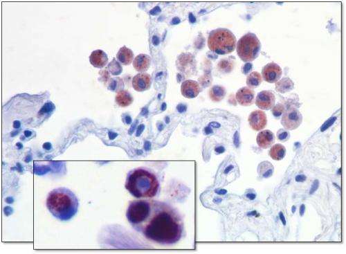
The team of researchers compared snap-frozen und HOPE-treated lung tissue from patients. In contrast to snap-frozen samples, HOPE fixation preserves the structure of tissues well and for example lung vesicles can be seen more clearly. The researchers then used mass spectrometry in order to characterise the proteins that are present in the tissue. The proteome derived from this study tells much about the health status of the tissue. The scientists went one step further and also investigated the so-called phospho-proteome, i.e. all protein molecules in the cell that are currently "switched on or off". To know which proteins are active contributes to the diagnosis of diseases and can help identify targets for new medications. The results are very promising: HOPE fixation does not only preserve the structure of the tissue but is just as well-suited for proteomics and phospho-proteomics as snap-freezing the tissue.
"Based on our results, we recommend HOPE as the fixation strategy for clinics and biobanks that are actively involved in improving diagnosis and therapies," says Jänsch.
The team of researchers applies this insight already in the research on legionnaire's disease, an infectious disease that is caused by bacteria and is associated with pneumonia. They maintain a close cooperation with Dr Torsten Goldmann, Research Centere Borstel, on this topic. "We already established an infection model for the human lung. We now know that HOPE makes this model also amenable to proteome and phospho-proteome analyses," says Prof Michael Steinert, who coordinates a project on this topic supported by the German Federal Ministry of Education and Research. "In the proteome analyses, we can already see some clear variations in the tissues of different donors and are starting to understand the individual infection process of legionnaire's disease better." HOPE thus lives up to its name and gives reason for hope in terms of new insights in the research, diagnosis and therapy of diseases.
More information: Olga Shevchuk, Nada Abidi, Frank Klawonn, Josef Wissing, Manfred Nimtz, Christian Kugler, Michael Steinert, Torsten Goldmann, Lothar Jänsch, HOPE-fixation of lung tissue allows retrospective proteome and phosphoproteome studies, Journal of Proteome Research, 2014, DOI: 10.1021/pr500096a


