Researchers identify brain mechanism for motion detection in fruit flies

A team of scientists has identified the neurons used in certain types of motion detection—findings that deepen our understanding of how the visual system functions.
"Our results show how neurons in the brain work together as part of an intricate process used to detect motion," says Claude Desplan, a professor in NYU's Department of Biology and the study's senior author.
The study, whose authors included Rudy Behnia, an NYU post-doctoral fellow, as well as researchers from the NYU Center for Neural Science and Yale and Stanford universities, appears in the journal Nature.
The researchers sought to explain some of the neurological underpinnings of a long-established and influential model, the Hassenstein–Reichardt correlator. It posits that motion detection relies on separate input channels that are processed in the brain in ways that coordinate these distinct inputs. The Nature study focused on neurons acting in this processing.
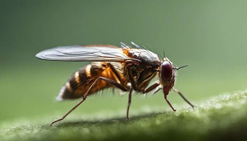
The researchers examined the fruit fly Drosophila, which is commonly used in biological research as a model system to decipher basic principles that direct the functions of the brain.
Previously, scientists studying Drosophila have identified two parallel pathways that respond to either moving light, or dark edges—a dynamic that underscores much of what flies see in detecting motion. For instance, a bird is an object whose dark edges flies see as it first moves across the bright light of the sky; after it passes through their field of view, flies see the light edge of the background sky.
However, the nature of the underlying neurological processing had not been clear.
In their study, the researchers analyzed the neuronal activity of particular neurons used to detect these movements. Specifically, they found that four neurons in the brain's medulla implement two processing steps. Two neurons— Tm1 and Tm2—respond to brightness decrements (central to the detection of moving dark edges); by contrast, two other neurons— Mi1 and Tm3—respond to brightness increments (or light edges). Moreover, Tm1 responds slower than does Tm2 while Mi1 responds slower than does Tm3, a difference in kinetics that fundamental to the Hassenstein-Reichardt correlator.
In sum, these neurons process the two inputs that precede the coordination outlined by the Hassenstein–Reichardt correlator, thereby revealing elements of the long-sought neural activity of motion detection in the fly.


