Image: How stem cells create just enough tissue
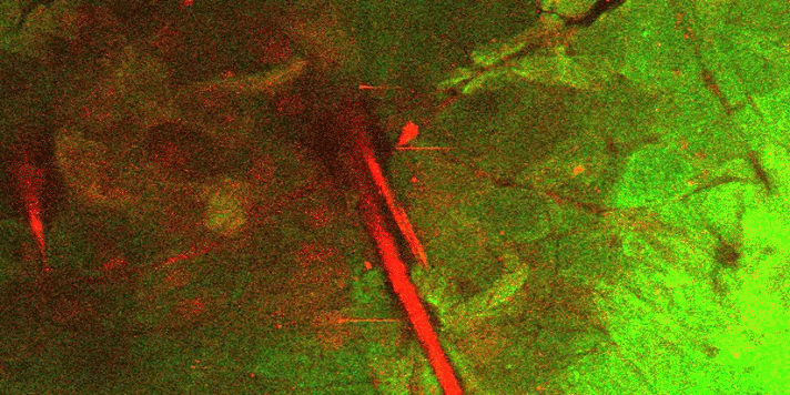
Scientists have long wondered just how stem cells generate just the right amount of tissue to perform a biological function. Using live imaging of skin of living mice, Yale scientists have shown that this balance, called homeostasis, is achieved in unexpected ways. In the accompanying movie, the researchers—led by Panteleimon Rompolas and Kailin R. Mesa in the lab of Valentina Greco—captured images of how stem cells (in red) at the bottom layer of epidermis (green) line up in columns and enlarge to replace cells lost at the top layer of skin.
The entire process is tightly coordinated in space and time in a process that challenges long-standing ideas of how stem cells function, note the researchers. The work was published online May 26 in the journal Science.
More information: P. Rompolas et al. Spatiotemporal coordination of stem cell commitment during epidermal homeostasis, Science (2016). DOI: 10.1126/science.aaf7012


