Visualizing how infection exacerbates lung disease

Chronic obstructive pulmonary disease (COPD) is a leading cause of death worldwide. The disease, characterized by constricted airways due to bronchitis and emphysema, is commonly seen in smokers but can also be brought on by environmental pollutants. The symptoms of the disease are known to be worsened by bacterial and viral infections of the airways, but exactly why remains largely unclear. A research team led by Naoyuki Taniguchi from the RIKEN Global Research Cluster has now developed a mouse model that allows COPD exacerbation to be studied in living animals.
Acute exacerbation of COPD as a result of infection of the airway is known to be a major cause of death in COPD patients. It is also known to accelerate the progression of the disease, but despite the disease's prevalence, surprisingly little is known about the mechanism of exacerbation or how to treat it. "Steroid therapy is common for COPD exacerbation, but its effectiveness is not clear," says Taniguchi. "It is reported to be effective in only 20% of patients, and sometimes causes serious side effects."
The development of a suitable animal model to specifically study COPD exacerbation is therefore of great interest to researchers looking for clinical treatments for these acute episodes.
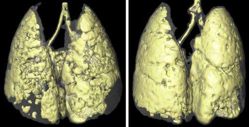
Satoshi Kobayashi and Reiko Fujinawa from Taniguchi's research group combined a simple mouse model of COPD exacerbation with an imaging technique called micro-computed x-ray tomography (micro-CT), which allowed them to monitor pathological changes by visualizing three-dimensional variations in tissue density. They produced their mouse model by exposing healthy mice to the chemical elastase, which results in the development of emphysema-like lung disease. Exacerbation of the disease was then triggered by exposing the mice to a single dose of lipopolysaccharide (LPS), a toxic component of bacterial cell walls that is known to cause inflammation.
The experiments showed that lung inflammation was exacerbated in mice exposed to LPS, with an associated increase in two different types of inflammatory cells and an increase in proinflammatory proteins and signalling molecules. Micro-CT imaging at 3 and 12 weeks following LPS exposure confirmed the acceleration of emphysema-like symptoms (Fig. 1).
The observed pathological changes mimic those found in human COPD exacerbation, meaning that this model could be used to help researchers gain a better understanding of the underlying mechanisms of COPD, and perhaps to develop new treatments for it. "Using this model mouse," says Taniguchi, "we are looking for biomarkers of the exacerbation process, and our final goal is to develop new therapeutics."
More information: Kobayashi, S., et al. A single dose of LPS into mice with emphysema mimics human COPD exacerbation as assessed by micro-CT, American Journal of Respiratory Cell and Molecular Biology, 3 July 2013. (DOI: 10.1165/rcmb.2013-0074OC). dx.doi.org/10.1165/rcmb.2013-0074OC


