Clearing up information about corneal dystrophies

Jayne Weiss, MD, Associate Dean of Clinical Affairs, Professor and Chair of Ophthalmology at LSU Health New Orleans School of Medicine, is the lead author of an editorial about inaccuracies in the medical literature that medical professionals rely upon to diagnose corneal dystrophies, as well as a free resource that provides correct information. The paper is published in the November 2018, issue of the American Journal of Ophthalmology.
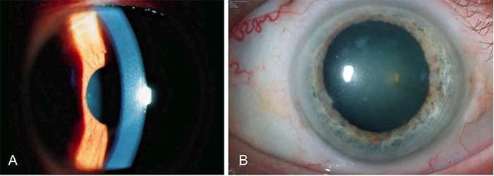
Corneal dystrophies comprise a group of relatively rare genetic eye disorders in which material accumulates in the cornea, the clear outer covering of the eye. This abnormal material can cloud the cornea, resulting in blurred or loss of vision in some patients. These disorders can affect both eyes, progress slowly and are hereditary. While there are some common characteristics, there are 22 distinct corneal dystrophies, and an accurate diagnosis is critical to proper treatment. Correctly characterizing a corneal dystrophy can be challenging, even for experts, much less clinicians who have never seen one. So clinicians frequently turn to the worldwide medical literature to learn about corneal dystrophies and often base their diagnoses upon information they find there.
Dr. Weiss and her colleagues write that publications on corneal dystrophies in the medical literature, however, can be confusing, contradictory or simply wrong. Errors arise from historical descriptions made before the invention of equipment like the slit lamp, confounding translations from scientific papers written in foreign languages, ignorance of previously published findings, misleading nomenclature, and difficulty in purging erroneous information from peer-reviewed journals and textbooks. As well, advances in molecular genetics and newly discovered information in other disciplines have expanded or changed our knowledge.
In 2005, Weiss created and continues to chair the International Committee for the Classification of Corneal Dystrophies (IC3D) to decrease published inaccuracies leading to confusion and provide clarity in diagnosing these disorders. Members from around the world are geneticists, pathologists and ophthalmologists specializing in corneal diseases with expertise in specific dystrophies.
A dynamic effort, IC3D periodically updates and revises its information. Its most recent article, a revision of its 2008 publication, was published in Cornea in 2015, available here.
This revision of the IC3D classification includes an updated anatomic classification of corneal dystrophies more accurately classifying one of the forms of corneal dystrophies that affect multiple layers rather than being confined to one corneal layer. Typical histopathologic and confocal images were also added to the corneal dystrophy templates.
"Our IC3D nomenclature for corneal dystrophies has become accepted internationally as the standard and is also used by the American Academy of Ophthalmology," notes Weiss. "I am proud that its easy accessibility by patients and clinicians alike has facilitated diagnosis and potentially treatment in individuals with these conditions."
More information: Jayne S. Weiss et al, The Corneal Dystrophies—Does the Literature Clarify or Confuse?, American Journal of Ophthalmology (2018). DOI: 10.1016/j.ajo.2018.07.047


