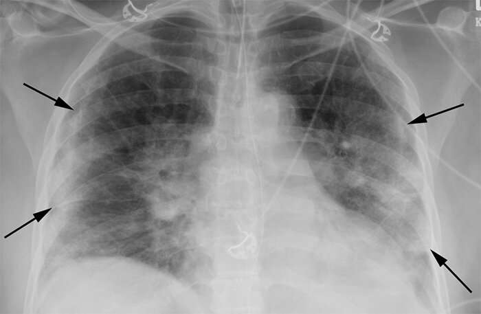
Radiologists find chest X-rays highly predictive of COVID-19

A team of LSU Health New Orleans radiologists investigated the usefulness of chest X-rays in COVID-19 and found they could aid in a rapid diagnosis of the disease, especially in areas with limited testing capacity or delayed test results. Their findings are published in Radiology: Cardiothoracic Imaging.
"In mid to late March of this year, when COVID-19 cases were spiking in New Orleans, we recognized an unusual pattern on chest X-rays that seemed to correlate with COVID positivity," notes David Smith, MD, Associate Professor of Clinical Radiology at LSU Health New Orleans School of Medicine. The radiologists conducted a retrospective study of nearly 400 persons under investigation (PUI) for COVID-19 in New Orleans. They reviewed the patients' chest X-rays along with concurrent reverse-transcription polymerase chain reaction (RT-PCR) virus tests. Using well-documented COVID-19 imaging patterns, two experienced radiologists categorized each chest X-ray as characteristic, nonspecific, or negative in appearance for COVID-19. The radiologists found a characteristic chest X-ray appearance is highly specific (96.6%) and has a high positive predictive value of 83.8% for SARS-CoV-2 infection in the setting of pandemic.
"The presence of patchy and/or confluent, band-like ground glass opacity or consolidation in a peripheral and mid-to-lower lung zone distribution on a chest radiograph is highly suggestive of SARS-CoV-2 infection and should be used in conjunction with clinical judgment to make a diagnosis," says Bradley Spieler MD, Associate Professor of Diagnostic Radiology and Vice Chairman of Research in the Department of Radiology at LSU Health New Orleans School of Medicine.
"The chest radiograph, while low in sensitivity, can indicate COVID-19 in patients whose radiographs exhibit characteristic COVID-19 findings, when used in concert with clinical factors," adds John-Paul Grenier, MD, an LSU Health New Orleans Radiology Resident. "While not a substitute for RT-PCR virus tests or Chest CT, radiographs could provide a rapid, cost-effective diagnosis of COVID-19 in a subset of infected patients during the COVID-19 pandemic. The utility of this technique is described in the context of known disadvantages of RT-PCR, considered the gold standard in COVID-19 diagnosis, and Chest CT, which is currently not recommended for COVID-19 diagnosis. "
"This discovery is useful to aid in diagnosis in the setting of pandemic spread of COVID-19, especially when adequate testing is lacking," says Dr. Smith.
"We believe this work has great potential to aid all health care providers in the fight against COVID-19," concludes Dr. Spieler.
Catherine Batte, MS, from the Department of Physics and Astronomy at Louisiana State University, also collaborated on the study.
"The COVID-19 pandemic has been especially tough on the people of New Orleans," declares Dr. Grenier. "We hope that the insights we've gained from studying this disease in our community can be used to help other communities across the globe."
More information: David L. Smith et al, A Characteristic Chest Radiographic Pattern in the Setting of COVID-19 Pandemic, Radiology: Cardiothoracic Imaging (2020). DOI: 10.1148/ryct.2020200280


