This article has been reviewed according to Science X's editorial process and policies. Editors have highlighted the following attributes while ensuring the content's credibility:
fact-checked
peer-reviewed publication
proofread
Lightweight, head-mounted microscope unveils brain oxygenation in freely moving mice

The brain consumes approximately 25% of the body's oxygen to fuel its neural activities, underscoring the importance of sufficient oxygen supply for maintaining normal cognitive operations. Consequently, monitoring cerebral oxygen levels is pivotal in assessing neurological health, identifying potential brain injuries, and enhancing patient outcomes in critical care settings.
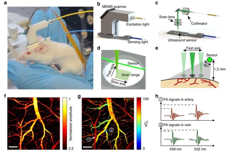
As reported in the latest issue of Light: Science & Applications, Prof. B. O. Guan and colleagues have introduced an innovative head-mounted microscope. This cutting-edge device enables continuous imaging of oxygenation in mice without restricting their movement.
This new microscope harnesses the capability of label-free photoacoustic imaging for cerebral oxygenation monitoring. It employs an optical fiber to guide and focus laser pulses into biological tissue. An additional fiber optic sensor is incorporated to detect the resulting ultrasound waves triggered by the laser.
Taking advantage of the distinct absorption spectra of oxygenated and deoxygenated hemoglobin, the team utilizes dual-colored laser pulses to measure blood oxygen saturation via a spectral unmixing process with single vessel resolution.
This miniature probe can be conveniently affixed to the head of a freely moving mouse, facilitating real-time imaging while observing the oxygenation and hemodynamics of individual blood vessels in the brain. For instance, the team showcased a short movie created through continuous brain imaging, illustrating the heightened oxygenation in the cortex during the transition from an anesthetized state to wakefulness.
Furthermore, they conducted meticulous investigations into the cerebrovascular response to acute hypoxia and hypercapnia challenges, all while the animals were in a freely moving state. This allowed the researchers to discern the distinct hemodynamic responses associated with different gas respirations and concentrations.
Notably, the imaging device was also utilized to study the impact of obesity on cerebral oxygenation in mice. The imaging findings revealed a significant reduction in the regulatory ability of cerebral vessels to external stimuli in obese mice. Importantly, this head-mounted imager effectively avoids the confounding effects of anesthesia, making it highly favorable and preferable for neuroscience and medical studies.
Cerebral oxygenation is intimately connected to neural activities through the mechanism of neurovascular coupling. Even a brief episode of brain hypoxia can result in irreversible damage and potentially lead to fatal outcomes.
The innovative device opens up new possibilities for detecting brain hypoxia and other injuries, while also offering a novel way to study both normal and pathological cerebrovascular functions. Furthermore, it provides invaluable insights into how various behaviors, including motion, influence brain function.
In addition, the imaging probe can be smoothly integrated with existing head-mounted neural imaging techniques, enabling the concurrent investigation of cerebrovascular and neurological activities.
This multifaceted device shows promise for studying the neural mechanisms behind social behaviors, as well as neurodegenerative disorders like Alzheimer's disease that disrupt neurovascular couplings. The photoacoustic fiberscope demonstrates potential for a broad spectrum of applications, extending beyond neuroscience research.
More information: Xiaoxuan Zhong et al, Free-moving-state microscopic imaging of cerebral oxygenation and hemodynamics with a photoacoustic fiberscope, Light: Science & Applications (2024). DOI: 10.1038/s41377-023-01348-3


