This article has been reviewed according to Science X's editorial process and policies. Editors have highlighted the following attributes while ensuring the content's credibility:
fact-checked
peer-reviewed publication
trusted source
proofread
Feature-domain Fourier ptychographic microscopy promotes pathological screening and analysis

Pathological analysis has been the gold standard of disease detection, especially in tumor diagnosis. Traditional digital pathology relies on high-precision movement of a high numerical aperture (NA) objective to obtain the entire field of view (FOV) for a tissue slide, whose high cost severely hinders its widespread applications.
Fourier ptychographic microscopy (FPM) digitalizes high-resolution images with large FOV without mechanical scanning, but suffers from vignetting artifacts, and becomes particularly susceptible to systematic errors.
Therefore, the application of FPM in digital pathology is limited to scientific research and has not been widely accepted for more than a decade.
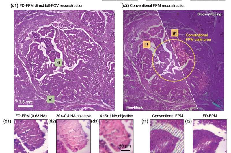
Recently, a research team led by Associate Prof. Pan An from Xi'an Institute of Optics and Precision Mechanics (XIOPM) of the Chinese Academy of Sciences (CAS) reported a feature domain-based FPM (FD-FPM) computational framework to realize non-blocked full-FOV reconstruction.
The study was published in Optica and selected as the cover story.
In their study, researchers theoretically pointed out that the fundamental reason for vignetting artifacts and sensitivity to systematic errors is the mismatch between forward model predictions and practical measurements.
Different from traditional methods focusing on the image itself, the reported FD-FPM switches to optimizing the feature of images. During the iterations, the updated target is transformed from traditional complex amplitude to complex gradient of images.
Such design plays an essential role in bridging the gap between theoretical model and experimental data. Intensive simulations and experiments have been conducted, and the results verified that FD-FPM enables elimination of vignetting artifacts at the edge of images without block operation and exhibits impressive robustness to various systematic errors (positional shifting of light source, uneven illumination intensity, etc.) and noise signals without data preprocessing.
Benefitting from the reported framework, the research team applied FPM to a self-developed whole slide imaging (WSI) platform and realized full-color imaging with 4.7mm diameter FOV at the highest resolution of 336 nm. This FPM-WSI instance demonstrated automatic high throughput imaging with low cost, and it will offer a turnkey solution to transform the high-quality WSI platform into one that can be made broadly available and utilizable.
The reported method is expected to break the bottleneck that has long been constraining the development of FPM since its invention in 2013, making FPM more broadly accepted and utilized in the field of biomedical research and clinical applications, according to experts in this area.
More information: Shuhe Zhang et al, FPM-WSI: Fourier ptychographic whole slide imaging via feature-domain backdiffraction, Optica (2024). DOI: 10.1364/OPTICA.517277


