This article has been reviewed according to Science X's editorial process and policies. Editors have highlighted the following attributes while ensuring the content's credibility:
fact-checked
trusted source
proofread
New study shows promising diagnosis of multiple sclerosis from images of the eye

Researchers at Durham University, UK and Isfahan University of Medical Sciences, Iran have developed an innovative approach to diagnosing multiple sclerosis using advanced eye imaging techniques.
The work is published in the journal Translational Vision Science & Technology.
This groundbreaking method could revolutionize how multiple sclerosis is detected, offering a faster, less invasive, and more accessible alternative to current diagnostic procedures.
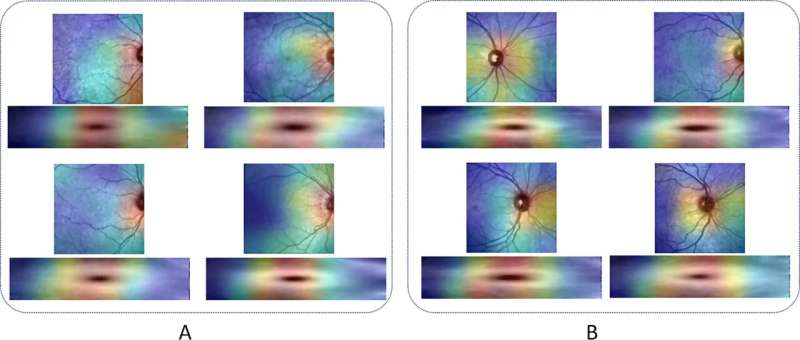
The study, led by Dr. Raheleh Kafieh of Durham University, integrates two types of eye scans: optical coherence tomography (OCT) and infrared scanning laser ophthalmoscopy (IR-SLO).
By training computer models with large numbers of these eye scans, the researchers have created a powerful diagnostic tool that can identify multiple sclerosis with remarkable accuracy.
What sets this approach apart is its ability to detect subtle changes in the eye that are often indicative of multiple sclerosis. The eye, being directly connected to the brain, can reveal early signs of neurological damage that might otherwise go unnoticed.
By training computers to recognize hidden patterns and abnormalities in eye images, this method offers the potential for earlier diagnosis and better management of multiple sclerosis symptoms.
The results of the study are impressive, with the computer model correctly identifying multiple sclerosis in 92% of cases during initial tests.
Even more encouraging, the system maintained a strong 85% accuracy when tested on a different set of data from other hospitals and populations, demonstrating its reliability and potential for widespread use.
Lead author of the study, Dr. Raheleh Kafieh of Durham University's Engineering department emphasized the significance of these findings and said, "Incorporating all available medical imaging, including those with subtle changes that are difficult to discern through non-computerized diagnosis, is crucial for achieving more reliable diagnoses and improving patient outcomes."
This innovative approach could have far-reaching implications for patients and health care providers alike. Early and accurate diagnosis of multiple sclerosis can dramatically impact the quality of life for those affected, potentially slowing the progression of the disease and improving overall outcomes.
Moreover, the non-invasive nature of eye scans makes this method more comfortable for patients and easier to implement in various health care settings, including high street opticians.
This novel eye imaging technique not only promises to enhance multiple sclerosis diagnosis but also opens doors for similar applications in other neurological conditions such as Alzheimer's and Parkinson's disease.
As this technology continues to develop, it could pave the way for more accessible and reliable diagnostic tools in everyday health care, ultimately leading to better patient care and outcomes.
More information: Roya Arian et al, SLO-Net: Enhancing Multiple Sclerosis Diagnosis Beyond Optical Coherence Tomography Using Infrared Reflectance Scanning Laser Ophthalmoscopy Images, Translational Vision Science & Technology (2024). DOI: 10.1167/tvst.13.7.13


