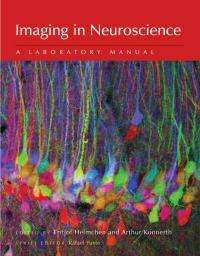
Cutting-edge imaging techniques for neuroscientists available in latest laboratory manual

Neuroscientists have long pioneered the use of new visualization techniques. Imaging in Neuroscience: A Laboratory Manual continues that tradition by presenting an outstanding collection of methods for visualizing the nervous system. It is the third volume in a series of imaging manuals published by Cold Spring Harbor Laboratory Press.
"This series of manuals originated in the Cold Spring Harbor Laboratory course on Imaging Structure and Function of the Nervous System, taught continuously since 1991," notes Rafael Yuste, the Series Editor. "Since its inception, the course quickly became a "watering hole" for the imaging community and especially for neuroscientists and cellular and developmental neurobiologists, who are traditionally always open to microscopy approaches."
In recent years, the ability to directly observe neural events at various spatial and temporal scales has enormously expanded because of the development of new imaging technologies and novel functional indicators. This manual provides the foundational methods for imaging nervous system cells and presents the latest techniques for visualizing cells, synapses, neuromolecules, circuits, and brain function in health and disease.
The manual is organized into eight sections that are roughly ordered according to spatial level of neural organization. Section 1 introduces molecular tools for labeling receptors and cells and for optical readout and control of neural activity. In Section 2 these tools are applied to the study of axonal propagation of excitation and synaptic transmission. Section 3 focuses on the integration of synaptic inputs in neuronal dendrites. Sections 4 and 5 provide tools for investigating functional properties of neurons and neural circuits in brain slice preparations (in vitro) and in living animals (in vivo), respectively. Section 6 looks at the dynamic properties of various glial cells, an important area of neuroscience. Section 7 presents new methods for performing high-resolution imaging studies of the brain during behavior. Finally, Section 8 provides examples of imaging studies in various animal models of disease.
More information: This new manual is the third volume in the imaging series that began with Imaging: A Laboratory Manual(www.cshlpress.com/link/imagingp.htm), released last November, followed by Imaging in Developmental Biology: A Laboratory Manual(www.cshlpress.com/link/imagingdevbiop.htm), released in January 2011.


