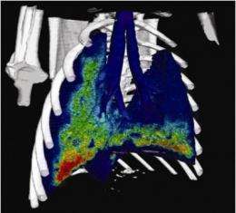
Viewing the lungs in 4D

(Medical Xpress) -- A new lung imaging method has the potential to revolutionise the study of lungs in both normal and diseased states.
Researchers from Monash University have developed a 4D dynamic functional lung imaging method, which allows them to study breathing lungs in unprecedented detail.
The method, developed by Faculty of Engineering PhD student Stephen Dubsky, provides measurements of motion, expansion and flow at every point within the lung, during the entire breathing cycle.
The technique utilises a synchrotron; essentially a very bright, high quality X-ray, to gather high-speed video of the breathing lung. Further research could allow translation to clinical X-ray imaging hardware, which may provide new a new clinical capability for diagnosis and management of lung disease.
Lead researcher, Dr Andreas Fouras from the Faculty of Engineering said the work, recently published in Journal of the Royal Society Interface, could allow for better understanding into the regional consequences of lung disease.
“The lung is a highly dynamic organ, and functional imaging can provide the next step in our understanding of the structure-function relationship within the lungs,” Dr Fouras said.
“Currently, a clinician treating a patient with a respiratory condition only has the options of spirometry and CT scans available to them. Typically, a CT provides regional information on the structure of the lung, while spirometry provides functional information averaged over the whole lung.
“The 4D functional imaging capability we have developed provides the best of both worlds, resulting in a detailed map of lung function.”
The technique lends itself to evaluation of respiratory conditions in which there may be alteration in the compliance of lung, chest wall and diaphragmatic function, or airway flow patterns. In particular, it may provide new insights into the regional consequences of restrictive lung disease, chronic obstructive airway diseases, and asthma.
“We are working with clinicians, funding bodies and industry partners to provide further impact for this work,” Dr Fouras said.
“The use of this technology will aid in the development and testing of new drugs and delivery methods, while further development towards clinical application may lead to new pathways for the diagnosis and monitoring of treatments for a variety of lung diseases.”
The project is a collaboration between the researchers from the Faculty of Engineering, Professor Stuart Hooper from the Monash Institute of Medical Research and Dr Karen Siu from Monash Biomedical Imaging and the Australian Synchrotron.
The team is the first in Australia, and only the third outside of North America, to win a prestigious and highly competitive American Asthma Foundation Award for their work.
More information: doi: 10.1098/rsif.2012.0116


