Too much salt could potentially contribute to liver damage

A sprinkle of salt can bring out the flavor of just about any dish. However, it's well known that too much can lead to high blood pressure, a potentially dangerous condition if left untreated. Now scientists report a new animal study that found a high-salt diet might also contribute to liver damage in adults and developing embryos. It appears in ACS' Journal of Agricultural and Food Chemistry.
Our bodies need a small amount of salt—the U.S. government recommends one teaspoon per day if you are a healthy adult. Among other functions, the sodium ions from the savory mineral help regulate water movement within the body and conduct nerve impulses. But most Americans eat too much salt. Some research indicates that in addition to high blood pressure, overconsumption of sodium can damage the liver. Xuesong Yang and colleagues wanted to explore the potential effect at a cellular level.
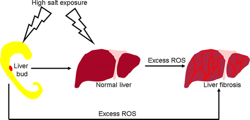
The researchers gave adult mice a high-salt diet and exposed chick embryos to a briny environment. Excessive sodium was associated with a number of changes in the animals' livers, including oddly shaped cells, an increase in cell death and a decrease in cell proliferation, which can contribute to the development of fibrosis. On a positive note, the researchers did find that treating damaged cells with vitamin C appeared to partially counter the ill effects of excess salt.
More information: Guang Wang et al. Liver Fibrosis Can Be Induced by High Salt Intake through Excess Reactive Oxygen Species (ROS) Production, Journal of Agricultural and Food Chemistry (2016). DOI: 10.1021/acs.jafc.5b05897
Abstract
High salt intake has been known to cause hypertension and other side effects. However, it is still unclear whether it also affects fibrosis in the mature or developing liver. This study demonstrates that high salt exposure in mice (4% NaCl in drinking water) and chick embryo (calculated final osmolality of the egg was 300 mosm/L) could lead to derangement of the hepatic cords and liver fibrosis using H&E, PAS, Masson, and Sirius red staining. Meanwhile, Desmin immunofluorescent staining of mouse and chick embryo livers indicated that hepatic stellate cells were activated after the high salt exposure. pHIS3 and BrdU immunohistological staining of mouse and chick embryo livers indicated that cell proliferation decreased; as well, TUNEL analyses indicated that cell apoptosis increased in the presence of high salt exposure. Next, dihydroethidium staining on the cultured chick hepatocytes indicated the excess ROS was generated following high salt exposure. Furthermore, AAPH (a known inducer of ROS production) treatment also induced the liver fibrosis in chick embryo. Positive Nrf2 and Keap1 immunohistological staining on mouse liver suggested that Nrf2/Keap1 signaling was involved in high salt induced ROS production. Finally, the CCK8 assay was used to determine whether or not the growth inhibitory effect induced by high salt exposure can be rescued by antioxidant vitamin C. Meanwhile, the RT-PCR result indicated that the Nrf2/Keap1 downsteam genes including HO-1, NQO-1, and SOD2 were involved in this process. In sum, these experiments suggest that high salt intake would lead to high risk of liver damage and fibrosis in both adults and developing embryos. The pathological mechanism may be the result from an imbalance between oxidative stress and the antioxidant system.


