Pelvic nerve visualizations reduce risk in surgery

Every year, thousands of patients suffer nerve damage in the pelvis during rectal surgery, leaving them with incontinence and sexual problems, for example. Noeska Smit has developed a method to make these nerves more visible to surgeons. She will be awarded a doctorate for her work on the subject at TU Delft on Monday, 31 October.
Background
The pelvis is anatomically complicated and certain details have not yet been fully mapped out. In just a small area of muscle and bone, there are numerous organs surrounded by blood vessels, nerves, lymphatic vessels and connective tissues. Surgeons operating on the pelvis need to have excellent knowledge of its microscopic anatomy. This can help prevent patients developing post-operative problems, such as incontinence or erectile dysfunction. In cancer surgery, good anatomical knowledge also helps ensure that the tumour is fully removed.
Model
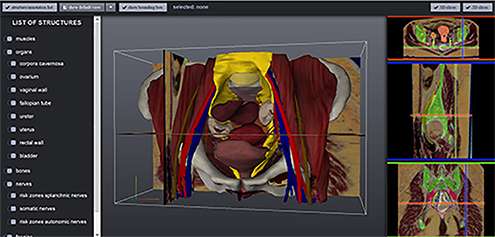
"This is why, in my research in the 'Computer Graphics & Visualisation' group, we worked together with the Leiden University Medical Center (LUMC) on a virtual 3-D model of the human pelvis, based on real anatomy",says Noeska Smit. Together with Annelot Kraima, who completed her PhD at Leiden University last year, she made a detailed virtual 3-D anatomical atlas combining all available and new information about the pelvis. "We then put this atlas to use both for educational purposes (to teach anatomy) and to improve pre-operative surgical planning."
Education
"For education, we created an online tool that enables anyone worldwide with an internet connection and a modern browser/PC to view our model. For instance, the tool has already been deployed in an anatomy MOOC (Massive Online Open Course) devised by Leiden University and it has been used by thousands of students already. The tool is free to consult anywhere in the world."

In practice
"As for surgical planning, it primarily concerns the treatment of rectal cancer, which affects around 4,000 people every year in the Netherlands alone. Some of the pelvic nerves are so small that they cannot be detected on an MRI scan and are hardly even visible to the surgeon during operations. As a result of this, the nerves are almost always damaged during surgery, often with serious consequences for patients (such as incontinence and sexual dysfunction)."
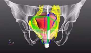
"Since our model shows these nerves, we can map them to an MRI of the person, so to speak, enabling us to chart these areas of nerves after all."
With the newly-developed tool, the areas of nerves can quickly be made visible. This enables the surgeon to see where the nerves are located relative to the tumour before the operation starts. This is set to prove especially useful for trainee surgeons, but also helps doctors to explain the risks involved in operations to patients.
More information: Tool 'The Online Anatomical Human': anatomy.tudelft.nl/


