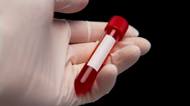
Cancers could be identified using blood tests

By enlarging molecules in the blood by 1,500 and marking them with a fluorescent, it may become easier to both identify signs of cancer and to find out if a treatment is effective. This is shown in a new thesis from Uppsala University.
"In the best of worlds, my method would make it possible to use a simple blood test to identify certain forms of cancer such as leukaemia or prostate cancer. It would make things easier for both hospital staff and patients, and in some cases also cheaper compared to the methods used today," says Liza Löf, researcher at the Department of Immunology, Genetics and Pathology at Uppsala University.
In her recently presented thesis, Liza Löf has further developed the proven molecular medicine method called proximity ligation essay (PLA) which was developed at Uppsala University. Previously this method has mostly been used in research studying how molecules interact with each other.
To study molecular events in blood, it is possible to use methods to identify and count small membrane-covered particles called vesicles. These vesicles are released from tissues and can transport molecules between cells, and they could be important biomarkers for cancers.
For instance, if a patient has a tumour somewhere in their body, this tumour may leak vesicles into the bloodstream where they can be identified and quantified. In the same way it is possible to find out if a patient is responding to cancer treatment. If the treatment is working, the cells of the tumour will break, producing many more vesicles in the blood than before the treatment was started.
Using this method to separate vesicles from different tissues in blood could simplify both diagnostics and patient follow-up.
"The problem up until now has been detecting these vesicles which are extremely small. By enlarging them they become easier to see, making the colour markings more effective and easy to work with. Instead of getting a fuzzy view of all vesicles we can now see them individually and connect them to possible diseases," says Liza Löf.
The type of fluorescent colouring used in the thesis is already being used to colour proteins.
Previous studies which have made use of this method have looked at fixed cells. What is new about Liza Löf's thesis is that she has studied certain cells in the blood. Studying vesicles is also new. By understanding where they come from it would be possible to find the possible location of the tumour in the body.
"The goal of my research has been to solve important problems that exist today on the treatment side, using our molecular methods. My studies have been conducted in close cooperation with physicians working with leukaemia patients."
More information: Applications of in situ proximity ligation assays for cancer research and diagnostics: uu.diva-portal.org/smash/record.jsf?pid=diva2%3A951029&dswid=-8769


