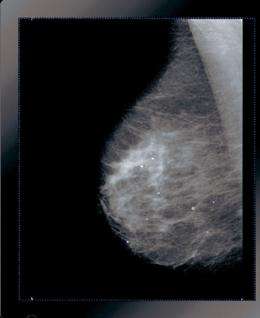
Breast cancer detection improved with image processing

Siemens researchers in Portugal hope to detect breast cancer more reliably in the future using a new statistical detection method. The digital image processing technique reveals tiny calcium deposits in the breast, which are a frequent indication of tumors. Previously only experienced radiologists could deduce the possible existence of such deposits from x-ray images. Computer-assisted detection of calcium deposits is very sensitive and can assist radiologists with their diagnosis. Comparisons with other methods have shown that the new method even permits conclusions to be drawn regarding the malignancy of the tumors associated with the deposits. The method is currently undergoing clinical trials.
Breast cancer is the most frequent cancer in women worldwide. The disease is curable if detected early enough. Screening is carried out on the basis of mammograms, which use x-ray images to reveal lumps in the breast. Calcium deposits can also indicate the existence of a tumor. However, the deposits are often only a few tenths of a millimeter in size and so deeply embedded in dense tissue that they are nearly undetectable in the images. Experienced radiologists know where and how to look for such signs. Digital image processing can assist with the correct interpretation of the images. However, the usual methods of noise suppression and image smoothing often also eliminate the tiny structures of the calcium deposits.
The Portuguese researchers at Siemens Healthcare take advantage of the fact that breast tissue displays self-similar properties, i.e., any excerpt, no matter how small or how highly magnified, resembles the complete tissue. The new recognition software thus reveals the calcium deposits as deviations from this self-similar structure, allowing the researchers to reliably visualize the tiny calcifications. They can also deduce the malignancy of the tumor from parameters such as the shape or distribution of the deposits. Comparisons with magnetic resonance imaging scans document that the tumors detected can be properly classified using the new method.


