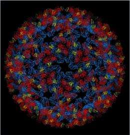
Elucidation of the 3D structure of Chikungunya virus

French researchers at the Pasteur Institute and the CNRS, in collaboration with the Synchrotron SOLEIL, have solved the three-dimensional structures of the glycoproteins that envelop the chikungunya virus. This discovery allows us to understand how this protein complex is activated in order to allow the virus to invade target cells. Activation is a key step in the viral life cycle, and its elucidation provides essential information for the development of antiviral strategies, for prevention and treatment.
The three-dimensional structures of the surface proteins which cover the Chikungunya virus particle, have been determined by the Structural Virology Unit of the Institut Pasteur (CNRS URA 3015), directed by Felix Rey, in collaboration with the Synchrotron SOLEIL PROXIMA1 beamline team, Institut Pasteur’s recombinant protein production platform (CNRS URA 2185) and Global Phasing Ltd., a company in Cambridge, UK.
The resolution of the 3D structure of these proteins and the complex they form, using crystallography and cryo-electron microscopy, shows their role in both the mechanism of invasion and that of the production of new viruses. Felix Rey's group has in fact determined the structure of two complexes, each with a specific role in different stages of the viral cycle: p62/E1 and E3/E2/E1, the second complex resulting from the maturation of the first.
In order to enter the target cell, the virus first uses E2 to attach itself to the cell membrane. The membrane of the cell then encloses the virus and seals it in vesicles that will transport the virus to successive cell compartments and direct it to the lysosome charged with dismantling it.
However the pH of these intermediate compartments, called endosomes, becomes progressively more acidic, resulting in the activation of E1. This protein will, in turn, ensure the fusion of the viral and endosomal membranes, so allowing the virus to release its RNA into the cell. This RNA is then taken up by the cell machinery.
Once the RNA is replicated, viral proteins organize to form new viruses capable of leaving the cell and infecting others. P62, insensitive to acidic pH, associates with E1 and allows the complex to migrate to the cell membrane. It is during this migration that p62 undergoes a maturation process that leads to the creation of proteins E2 and E3. The E3/E2/E1 complexes, thus formed, group together to create new viral particles budding from the surface of the infected cell in order to invade new cells.
Understanding these mechanisms is an important step in identifying therapeutic targets: it shows that stabilization of the E3/E2/E1 complex would stop the virus from invading the cell. The study also identified areas on E2 that recognize neutralizing antibodies, paving the way for new approaches to vaccines.
The chikungunya virus, transmitted to humans when bitten by mosquitoes of the genus Aedes, causes severe joint pain in patients. Today, medical treatment for infection is to prescribe anti-inflammatories and painkillers. The 2005-2006 epidemic in the Indian Ocean affected almost 2 million people in India and around 270,000 on the island of Reunion. A new outbreak of the disease occurred in Spring 2010 and the first two indigenous cases were reported in France in September 2010.
More information: Glycoprotein organization of chikungunya virus particles revealed by X-ray crystallography, Nature, 2010, 468(7324): 709–712 (published 2nd December, 2010).


