Breast cancer 'clearance' techniques suggested

AN investigation into the methods of checking that breast cancer is completely removed during surgery, has found some methods aren't affective at eliminating the need for a second surgery.
The review, published in the journal The Breast, examined the success rate of intraoperative assessment methods – frozen section, imprint/touch smear cytology and ultrasounds – used in breast cancer surgery.
Lead author and Curtin University's Kerryn Butler-Henderson says intraoperative methods help to determine how much of a tumour remains in the breast during the first operation.
"Where accurate, this means the tests not only reduce the need for a second operation but can potentially reduce the rate of recurrence or invasion," Ms Butler-Henderson says.
"Second operations are often required when the breast cancer is close or involved in one or more surgical margins.
"Research has shown that where an adequate margin has not been achieved there is an increased risk of recurrence or invasion."
She says it is important to ensure adequate margins have been obtained in surgery.
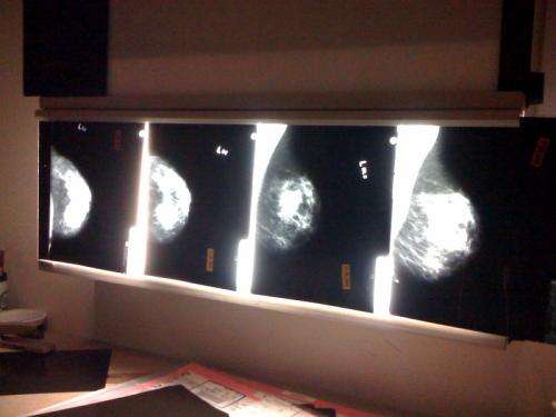
"The problem is this can be difficult to achieve as the surgeon only has mammography images to view and if the tumour is large enough they can palpate the tumour," she says.
"In many cases the patient will have a hookwire inserted to guide the surgeon but my epidemiological study has shown in ductal carcinoma in situ (DCIS) alone, 18 per cent of women [nearly one in five] will still need a second operation to obtain clearance."
To prevent patients having to come back for another operation, doctors use intraoperative methods to assess the area, checking that all infected tissue is removed.
"Although pathological methods, such as frozen section and imprint cytology performed well [in the studies reviewed], they added on average 20–30 minutes to operation times," Ms Butler-Henderson says.
"During imprint or touch smear cytology, [surgeons] 'rub' the margin surface onto a slide and look at that microscopically—the problem with this is not only does it take approximately 20 minutes, you cannot determine if there is a close margin, just if there is DCIS on the margin.
"An ultrasound probe allows accurate examination of the margins and delivers results in a timely manner, yet it has a limited role with DCIS where calcification is present and in multifocal cancer."
Ms Butler-Henderson instead recommends further research be undertaken in the use of digital mammography, radiofrequency spectroscopy, or optical coherence tomography.
"I have also been exploring, with success, the use of a positron emission tomography (PET) probe in surgery," she says.
More information: "Intraoperative assessment of margins in breast conserving therapy: A systematic review." Kerryn Butler-Henderson, Andy H. Lee, Roger I. Price, Kaylene Waring. The Breast - 27 January 2014 (10.1016/j.breast.2014.01.002)


