Researchers develop new animal model to study rare brain disease

Thanatophoric dysplasia (TD) is an intractable disease causing abnormalities of bones and the brain. In a recent study of ferrets, which have brains similar to those of humans, researchers using a newly developed technique discovered that neuronal translocation along radial glial fibers to the cerebral cortex during fetal brain development is aberrant, suggesting the cause underlying TD.
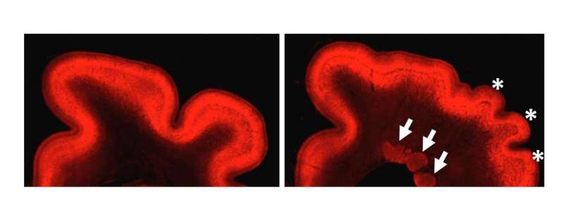
In TD cases, the limb and rib bones are shorter than normal, and brain abnormalities manifest, including polymicrogyria and periventricular nodular heterotopia. Previous research has determined that a gene, fibroblast growth factor receptor 3 (FGFR3), is responsible. However, as a result of TD rarity and the difficulty of obtaining brain samples from human patients, the pathophysiology of TD is largely unknown, and effective therapy has not been established.
The present research team of Kanazawa University generated an animal model of TD using ferrets that reproduces the brain abnormalities found in human TD patients. By using this animal model, the team elucidated the formation process of polymicrogyria, one of the abnormalities found in the TD brain. The team has also investigated the formation process of PNH, the other brain abnormality found in human TD patients.
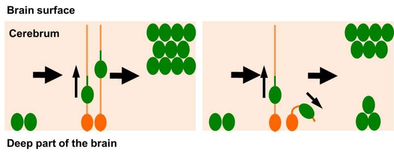
First, PNH was analyzed in terms of composing cell types to reveal that a large number of neurons but few glial cell exist in PNH. In a healthy brain, neurons are found in the cerebral cortex near the brain surface. The researchers believe that during fetal brain development, PNH formation might be induced by the inability of neurons to translocate themselves to the cerebral cortex. The researchers found that the spatial arrangement of radial glial cells was distorted; radial glial fibers are believed to serve as the "track" for neurons to translocate themselves. Thus, the distortion of radial glial fibers seems to be a reason for aberrant localization of neurons.

Research on abnormalities of bones in TD is progressing with iPS cells at Kyoto University, and it is expected that the whole aspect of TD with brain and bone abnormalities would be elucidated and that the therapeutic methods would be developed. The present study on PNH was only possible using the experimental technique for ferrets developed by the research team. This animal model technique could also contribute to studies of other neurological diseases that have been difficult to investigate with conventional model animals.
More information: Naoyuki Matsumoto et al, Pathophysiological analyses of periventricular nodular heterotopia using gyrencephalic mammals, Human Molecular Genetics (2017). DOI: 10.1093/hmg/ddx038


