SNMMI Image of the Year: PET and optical imaging for prostate cancer diagnosis and therapy

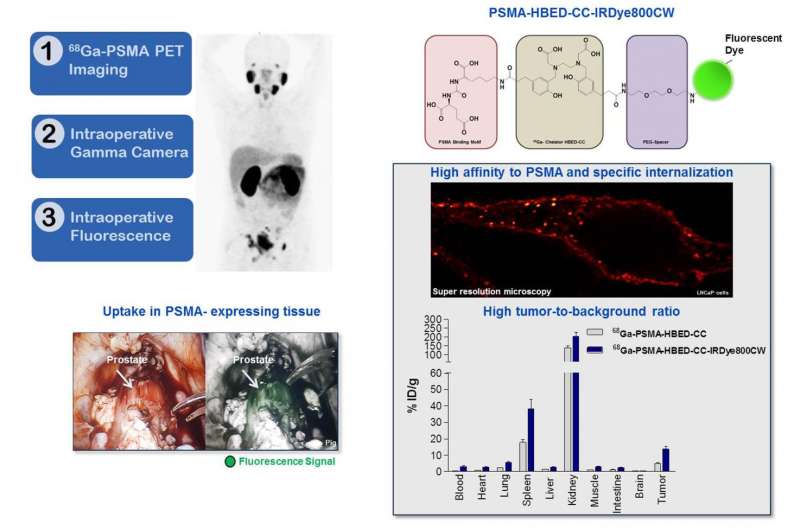
In the battle against metastatic prostate cancer, the removal of lymph node metastases using image-guided surgery may have a high clinical impact on outcomes. Researchers at the 2017 Annual Meeting of the Society of Nuclear Medicine and Molecular Imaging (SNMMI) demonstrated preclinically that dual-labeled PSMA-inhibitors based on PSMA-11 enhance preoperative staging, using PET/CT followed by fluorescence-guided surgery. The combined approach results in more accurate detection of PSMA-positive tumor lesions.
Each year, SNMMI chooses an image that exemplifies the most promising advances in the field of nuclear medicine and molecular imaging. The state-of-the-art technologies captured in these images demonstrate the capacity to improve patient care by detecting disease, aiding diagnosis, improving clinical confidence and providing a means of selecting appropriate treatments. This year, the SNMMI Image of the Year was chosen from more than 2,000 abstracts submitted to the meeting and voted on by reviewers and the society leadership.
The 2017 Image of the Year goes to a team of researchers from the German Cancer Research Center (DKFZ) and University Hospital in Heidelberg. The image clearly demonstrates how combining the advantages of 68Ga-PSMA PET and intraoperative gamma and fluorescence imaging results in better tumor identification before and during surgery.
"We are deeply honored to receive this award, and I would like to thank all team members who contributed to this interdisciplinary work," said Ann-Christin Baranski. "As resection of lymph node metastases has considerable impact on the outcome of metastatic prostate cancer patients, the aim of our study is to improve the intraoperative accuracy of detecting PSMA-positive tumor lesions."
"There has been a huge effort to improve care of prostate cancer patients using molecular imaging," stated Satoshi Minoshima, MD, PhD, chair of the SNMMI Scientific Program Committee and SNMMI vice president-elect. "The study presented by Ann-Christin Baranski clearly demonstrates that the combined PET imaging, gamma detection, and optical imaging can help not only pre-operative staging of the disease but also intra-operative guidance of metastatic lymph node dissection. We anticipate that such hybrid cancer detection methods will become prevalent in the near future and contribute significantly to the care and management of prostate cancer patients."
More information: "Preclinical evaluation of dual-labeled PSMA-inhibitors for the diagnosis and therapy of prostate cancer." SNMMI's 64th Annual Meeting, June 10-14, 2017, Denver, Colo.


