Researchers find new biomarker for active sarcoidosis

Low blood levels of immune cells called lymphocytes, in combination with higher levels of inflammation on PET/CT scans, are indicators of active sarcoidosis—an inflammatory disease that attacks multiple organs, particularly the lungs and lymph nodes—which disproportionately affects African Americans. The discovery by researchers at the University of Illinois Chicago could help guide disease treatment. Their findings are published in the journal Frontiers in Medicine.
The researchers were looking for biomarkers—both in the blood and in PET/CT scan findings—that would cluster patients into groups based on disease activity. Symptoms of sarcoidosis can come and go and the disease can switch from active—where granulomas form and enlarge—to inactive states where the disease is dormant and symptoms may lessen. There are few tests that can determine whether sarcoidosis is in the active or inactive stage.
"Having a biomarker signature that indicates active inflammation could be very useful in helping guide treatment and for helping us understand the underlying mechanisms associated with the disease," said Dr. Nadera Sweiss, professor of medicine and chief of the department of rheumatology in the UIC College of Medicine and corresponding author of the paper.
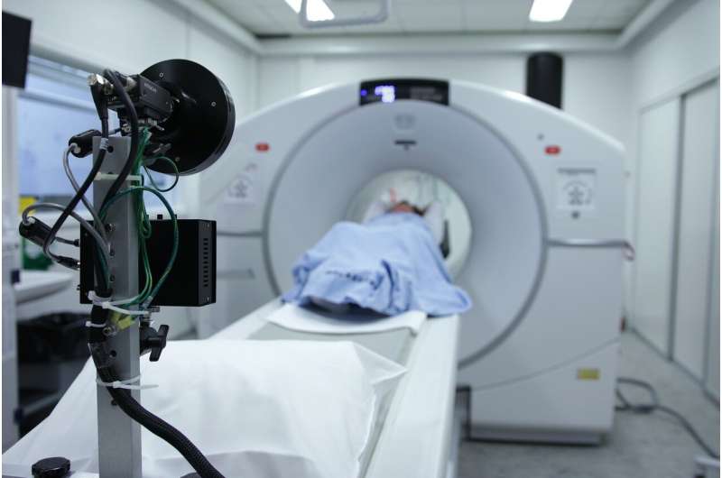
The researchers looked at the medical records of 58 patients—mostly African American—who were diagnosed with sarcoidosis based on biopsy results and who also underwent PET/CT scanning at the Bernie Mac Sarcoidosis Translational Advanced Research (STAR) Center at UI Health, the academic health enterprise of UIC. These patients all had blood tests that included lymphocyte counts. PET/CT scans provide images that show where active inflammation is occurring in the body.
The researchers found three distinct patient segments that could be grouped based on shared characteristics from lymphocyte counts and PET/CT scan findings. The first group included patients with chronic but quiescent—dormant—disease. Group two included mostly African American patients with some markers of general inflammation as seen on PET/CT scans who also had advanced pulmonary disease, and group three included predominantly Caucasian patients with reduced lymphocyte counts and acute disease. In contrast to group one, groups two and three had elevated levels of inflammation as picked up by the PET/CT scans. The researchers also found that lymphocyte levels could predict levels of inflammation seen on PET/CT scans, with lower lymphocyte levels associated with greater levels of inflammation as picked up on the scans.
"We found that active inflammation, as shown in PET/CT scans, was associated with active disease states," said Dr. Christian Ascoli, a resident in the departments of internal medicine, pulmonary and critical care at UIC, and an author on the paper. "Lymphocyte levels were lower in patients with more inflammation, indicating that lymphocyte levels are also a marker of active disease."
Using measurements of blood lymphocyte levels in combination with PET/CT scans could help guide treatment, Sweiss explained, since treatment differs for active versus inactive disease states.
"Our finding represents another tool in our toolbox for guiding treatment of this very variable disease, and helping us learn more about its underlying biology," she said.
More information: Christen Vagts et al, Unsupervised Clustering Reveals Sarcoidosis Phenotypes Marked by a Reduction in Lymphocytes Relate to Increased Inflammatory Activity on 18FDG-PET/CT, Frontiers in Medicine (2021). DOI: 10.3389/fmed.2021.595077


