January 17, 2022 feature
Enthesis strength, toughness, and stiffness: Comparing tendon insertions with varying bony attachment geometries
 for supraspinatus, Achilles, and patellar tendon with corresponding contrast-enhanced images of murine rotator cuff (supraspinatus tendon), ankle (Achilles tendon), and knee (patellar tendon) joints held in flexion, straight, and in extension. Green dashed lines outline tendons at each position. HH, humeral head; G, glenoid. Credit: Royal Society Interface, DOI: 10.1098/rsif.2021.0421")
While tendons of the body differ dramatically in their function, mechanics, and the range of motion, they all connect to bone via an enthesis—a specialized interface that integrates tendon into bone. Effective force transfer at the enthesis can allow joint stability and mobility, with strength and stiffness due to a fibrous architecture. However, the mechanisms of enthesis toughness can arise across tendons with different load orientations, which remain to be understood. In this work, Mikhail Golman, and a team of scientists at the Columbia University, New York, Missouri University, MO and the Washington University, St. Louis, U.S., performed simultaneous imaging of the bone and tendon in enthesis to represent variations of tendon-to-bone insertion. They extended the mathematical model to account for variations in insertion and bone geometry and tested the hypothesis that the toughness across a range of tendon enthesis could be observed via different interactions between fiber and bone architectures. The toughness of the model developed here arose from fiber re-orientation, recruitment and rupture mechanisms, mediated by interactions between fibers at the enthesis and bony ridge around it. In application to tendons, some were characterized as either energy storing or positional. The results provided insight into methods for surgical repair of tendon-to-bone attachments in orthopedics medicine, and the effects of attaching highly dissimilar materials, for bone tissue engineering. The work is now published in the Journal of the Royal Society Interface.
Muscle architecture
Joint stability and mobility can be ensured by muscle forces that transfer across the tendon-to-bone attachment via a transitional tissue known as the tendon enthesis. This is applicable across an entire range of tendons including 'positional' and 'energy storing' tendons with compositional differences for specific functions. In order to understand the effect of the simple fibrous architecture and motif, and see how the tailed bone ridge motif could provide different entheses with resilience at diverse insertion sites, and loading directions, the scientists performed imaging and modeling analyses. In this work, Golman et al. determined the roles of architectural and positional factors of enthesis toughness. To accomplish this, they applied mercury (III) chloride contrast micro-CT imaging to murine patellar, supraspinatus and Achilles tendons to identify qualitative differences in the nature of fibrous insertions and the bone surface that interact with such muscles. Golman et al. examined a model of these data to show that the interactions mediated strength, stiffness and toughness across a range of loading orientations. The outcomes provided insight to show how a single motif in insertion anatomy can provide insight to a range of insertions to describe the bony anatomy during fiber recruitment and tendon enthesis mechanics.
After harvesting the murine patellar, supraspinatus and Achilles' tendon-to-bone attachment units from healthy murine, the scientists submerged enthesis samples in mercury chloride for 24 hours and visualized them using X-ray computed tomography. The team then used a mathematical model based on bioimaging the fibrous architecture and the bony anatomy of the enthesis to predict variations of the toughness of the tendon enthesis with loading direction, fiber recruitment and interactions between the tendon and bony ridge. Golman et al. defined toughness as the energy required to displace the muscle-part of an idealized tendon. The simulations revealed distinct mechanical responses across a range of physiologically relevant loading directions. They then then plotted the relationship between fiber engagement and displacement to represent the percentage of fibers engaged at a given displacement. The model reproduced many trends and variations in mechanical behavior as seen in tendon enthesis mechanical testing experiments.
 Coronal section of high-resolution contrast-enhanced microCT image of a murine supraspinatus tendon attached to the humeral head. (b) Tendon enthesis fibers are shown in green, the semi-elliptical bone (of major axis A0 and minor axis B0) is shown in grey and the unit vector, ê, represents the direction of loading at an angle θ relative to the horizontal unit vector î. Fiber kinematics were followed to track the region over which each fiber contacted its neighbor (the region between angles Φ1 and Φ2, at points a distance r1 and r2, respectively, from the origin shown). These positions changed as the distal end of the tendon was displaced (that is, as r3 increased). The black arrow represents the direction of loading. (c) At rest, the length of the outer fibers is larger than the length of the inner fibers. When a fiber is engaged and oriented so that it contacts the bone ridge, it tightens until it reaches the tangent to the humeral head curvature (blue arrow) and the tangent point to the tendon fiber (purple arrow). (d) To approximate the distribution of fibers at the enthesis, with middle fibers being more numerous and storing more energy, fiber density was varied across the attachment. Specifically, (d, top) the distance between fibers was increased at the outer edges and (d, bottom) fiber stiffness K was increased for middle fibers. Credit: Royal Society Interface, DOI: 10.1098/rsif.2021.0421")
The influence of fiber packing density on enthesis mechanics
To understand the effect of fiber density on tendon enthesis mechanics, Golman et al. next varied fiber packing in the model to adjust the ratio between fiber spacing and thickness. The simulations revealed how the spacing between fibers affected the normalized strength, stiffness and toughness, where the effect depended on the joint position. When the tendon enthesis was loaded at 120 degrees, the normalized strength and toughness were lowest, despite the degree of fiber packing. When fibers were tightly packed, the strength did not depend on the loading angle. The team next identified how the enthesis of tendons commonly classified as 'energy-storing' prioritized toughness compared to strength, while those commonly classified as 'positional' demonstrated consistent stiffness. Golman et al. concluded the experiments by describing the influence of bony ridges at the enthesis to govern fiber recruitment and ultimately regulate enthesis mechanics.
-
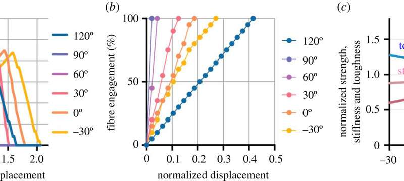
Positional-recruitment simulation results. (a) Normalized strength–displacement plot from simulations reproduced many trends and variations of loading angle-dependent mechanical responses seen in ex vivo experimental data. (b) Number of fibers engaged (recruited) with respect to displacement dependent on loading angle. (c) Normalized strength, stiffness, and toughness responses for tendon enthesis simulated to be loaded in range of loading directions. Simulations were run with 20 fibers (N = 20), round bone ridge (A0 = B0), fiber thickness of w = 10 µm, fiber spacing s/w = 1.5 and fiber failure stretch of λu = 1.2. Strength, stiffness and toughness were normalized against idealized scenario where tightly packed fibers (s/w = 1) were pulled to failure uniaxially at 90°. Credit: Royal Society Interface, DOI: 10.1098/rsif.2021.0421 -
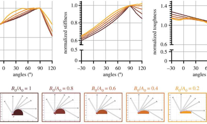
Anatomical bone ridge flattening on tendon enthesis mechanics. Simulations were run with 20 fibers (N = 20), fiber thickness of w = 10 µm, fiber spacing s/w = 1.5, and a variable aspect ratio for the cylindrical bone ridge. Fiber failure stretch was set to λu = 1.2. Initial conditions were set as for energy-storing tendons. Strength, stiffness, and toughness were normalized against idealized scenario where tightly packed fibers (s/w = 1) were pulled to failure uniaxially at 90°. Credit: Royal Society Interface, DOI: 10.1098/rsif.2021.0421
Outlook
In this way, Mikhail Golman and colleagues investigated how tendon enthesis achieved a wide range of functions by developing a position dependent fiber recruitment model, which described the range of tendon entheses and bone attachment geometries. The simulations highlighted the toughening mechanisms across a range of joint orientations and revealed tendon enthesis toughness across a range of joint motion, driven by fiber architecture, including fiber orientation, recruitment, and rupture. The simulations highlighted the entheses of tendons commonly characterized as 'energy-storing,' which prioritized toughness above strength, while those characterized as 'positional' prioritized consistent stiffness. The bony anatomy of tendon attachment dictated fiber recruitment patterns and enthesis mechanics to reveal toughening mechanisms that protect the tendon-bone interface from injury, important in clinical orthopedics. The team also identified several parameters that are important for the attachment of architecture materials, useful for applications in bone tissue engineering.
More information: Mikhail Golman et al, Enthesis strength, toughness and stiffness: an image-based model comparing tendon insertions with varying bony attachment geometries, Journal of The Royal Society Interface (2021). DOI: 10.1098/rsif.2021.0421
L. Rossetti et al, The microstructure and micromechanics of the tendon–bone insertion, Nature Materials (2017). DOI: 10.1038/nmat4863
© 2022 Science X Network


