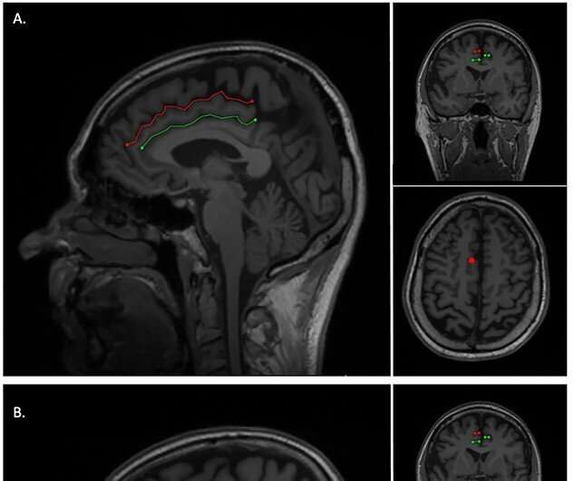
Brain folds formed during fetal stage could affect onset of dementia

Frontotemporal dementia begins relatively early compared to other forms of dementia. We still know relatively little about what causes the disease, however, new research from Lund University in Sweden shows that the folds in the brain that are formed in the latter stages of pregnancy could have an impact on the age at which the disease takes hold.
Those affected by frontotemporal dementia usually experience an early onset, often while still of working age. In addition, the progression of the disease is rapid and the average survival time after diagnosis is eight years.
"There is no cure, or treatment to slow progression. Therefore, any new knowledge of the disease is valuable," says Alexander Santillo, Associate Professor of Psychiatry at Lund University and consultant psychiatrist.
In a study with 307 participants (27–87 years of age), of which 92 were suffering from Alzheimer's disease and 105 from frontotemporal dementia (also known as frontal lobe dementia) and a control group of 110 people, Alexander Santillo and his colleagues examined the patients' brains with MRIs. They discovered that the brain folds that form during fetal development may have an impact on the age at which individuals begin to suffer from frontotemporal dementia. In those suffering from Alzheimer's disease, this did not seem to have an impact on the illness.
"The specific area we have studied is called the cingulate gyrus, which is situated in the frontal lobe of the brain. The study participants who had an extra fold in this area in the right cerebral hemisphere developed symptoms of frontotemporal dementia an average of 3 years later than individuals without this fold," says Luke Harper, neurology resident, doctoral student at Lund University and first author of the study published in Cerebral Cortex.
The researchers behind the study say that the findings contribute to a dramatic increase in understanding of the disease. It was already known that genetics has a bearing on whether you will develop dementia, however, the link to how the brain develops in the fetal stage had not previously been made.
"During this period, the brain is particularly sensitive to environmental factors. In the past, it was not possible to make connections between fetal development and diseases that affect the brain 60–70 years after birth. Therefore, we believe the findings may also be of relevance to other neurological diseases," explains Alexander Santillo.
Why this anatomical variation seems to affect the right cerebral hemisphere but not the left is not entirely clear, however, those suffering from frontotemporal dementia experience behavioral changes involving the right side of the brain. It is also already known that the anterior part of the cingulate gyrus in the cerebral cortex is affected by this specific type of dementia.
"We know that some people are affected by frontotemporal cognitive disease due to a genetic mutation, however, for the vast majority of people the cause is unknown, as are the factors that influence the progression of the disease. Our findings also indicate that variations in brain folds need to be taken into account when developing new treatments. However, our findings need to be confirmed in other studies first," says Alexander Santillo.
Luke Harper has begun work to confirm the findings in collaboration with American and Dutch research teams. The aim is to also look at whether the folds in this region of the brain affect the rate of progression of the disease and how long the patients survive after a diagnosis.
More information: Luke Harper et al, Prenatal Gyrification Pattern Affects Age at Onset in Frontotemporal Dementia, Cerebral Cortex (2021). DOI: 10.1093/cercor/bhab457


