August 16, 2022 report
Evidence of SARS-CoV-2 virus infecting astrocyte cells in the brain

A team of researchers affiliated with several institutions in Brazil has found evidence of the SARS-CoV-2 virus infecting astrocyte cells in the human brain. In their paper published in Proceedings of the National Academy of Sciences, the group describes their study of the brains of people who had died from COVID-19 and what was found.
From the earliest days of the pandemic, a large number of infected people have been complaining of neurological problems, such as brain fog, headaches and trouble paying attention. Because of that, medical scientists have been studying the problem to learn more about how the SARS-CoV-2 virus might infect the brain.
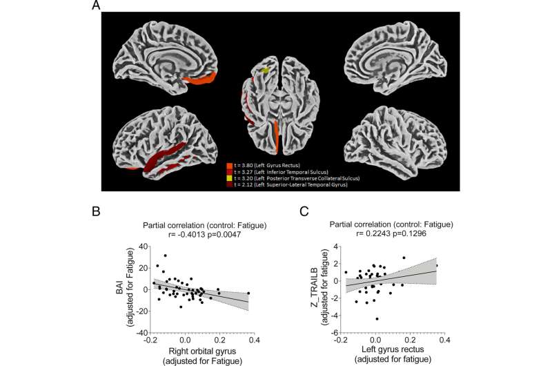
The work by the team on this new effort began with a study of 81 people who had been infected but had not died, or even been hospitalized. In comparing the group to a control group that had not been infected, the researchers found them to exhibit more symptoms of depression and anxiety. The researchers noted that such symptoms are typical of problems in the orbitofrontal cortex.
The researchers next dissected the brains of 26 people who had died from COVID-19, focusing specifically on the orbitofrontal cortex. In so doing, they found the virus present in astrocytes in five of them, though they note it was possible that the virus had been missed in the brains of some of the other dead patients.
Astrocytes exist in the brain but are not nerve cells; they are star-shaped glial cells that provide support for the neurons—they make and transport food to them. In taking a close look at the viruses that had infected the astrocytes, the researchers found they produced a protein that changed the behavior of the astrocytes—they made less lactate, which is food for neurons.
The researchers then looked into how the virus was able to infect the astrocytes. They found that spike proteins on the SARS-CoV-2 virus targeted different receptors than those that are targeted in the lungs, allowing them to bond with astrocytes. The end result, the researchers found, was death of neurons due to the inability of infected astrocytes to feed them.
More information: Fernanda Crunfli et al, Morphological, cellular, and molecular basis of brain infection in COVID-19 patients, Proceedings of the National Academy of Sciences (2022). DOI: 10.1073/pnas.2200960119
© 2022 Science X Network


