Team uses open-source toolbox for harmonized analysis of clinical angiography images

Dr. Danuta Sampson, a visiting senior researcher at the University of Surrey, with her colleagues, has embraced open research principles to support the discovery of novel biomarkers aimed at reducing the mortality of vascular disease, the leading cause of death worldwide.
Accurate assessment of the microvasculature (the smallest vessels in the human body) could identify biomarkers that lead to a decline in vascular disease mortality. Optical coherence tomography angiography (OCTA) is a non-invasive modality capable of imaging microvasculature in the human retina and in the skin. OCTA studies have been performed using many different lab-based and commercial clinical instruments, imaging protocols, data analysis methods, and metrics, often applied inconsistently and only partially reported, resulting in a confusing picture that represents a major barrier to progress in OCTA biomarker discovery.
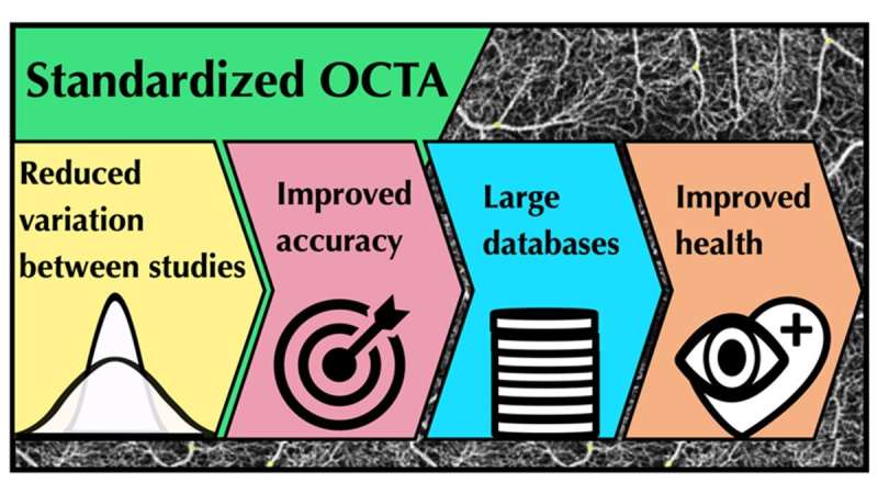
Open data and software sharing, and cross-comparison and pooling of data from different studies are rare. This lack of good open science practice has impeded building the large databases of annotated OCTA images of healthy and diseased retinas and skin that are necessary to study and define characteristics of specific conditions. The research team's project aims to remove this barrier by introducing and making publicly available software that enables OCTA image analysis in a transparent, harmonized and ultimately standardized way.
The team used skin OCTA images to develop and optimize a toolbox for OCTA image analysis. They validated the optimized software using OCTA images from their own and different commercial and non-commercial instruments and samples.
The researchers created an integrated MATLAB—ImageJ toolkit (OCTAVA—OCTA Vascular Analyser) with a user-friendly interface for processing and analysis of OCTA images. Quantitative results from various OCTA images showed that OCTAVA can accurately and reproducibly determine metrics for characterization of the microvasculature. The OCTAVA software is in open repositories to facilitate transparent and efficient collaboration. Source code is available on Github. A compiled MATLAB app or standalone version of the software—for a user without a MATLAB license, is available at Sourceforge or on request. All software and source code are open source under the MIT license.
There are no large-scale OCTA datasets yet widely available. Making OCTAVA open access means that it can be further validated via international laboratory and clinical research communities and eventually become standardized software for OCTA data analysis. This would enable building large cross-institution normative databases of the microvascular system in health and disease.
Such large data sets will enable defining the most sensitive biomarkers to distinguish between health and vascular disease.
Related research was published in Light: Science & Applications and PLOS ONE.
More information: Danuta M. Sampson et al, Towards standardizing retinal optical coherence tomography angiography: a review, Light: Science & Applications (2022). DOI: 10.1038/s41377-022-00740-9
Gavrielle R. Untracht et al, OCTAVA: An open-source toolbox for quantitative analysis of optical coherence tomography angiography images, PLOS ONE (2021). DOI: 10.1371/journal.pone.0261052


