A window into the changing tumor microenvironment

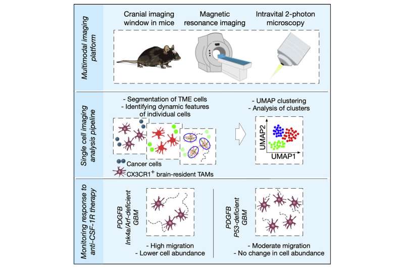
Researchers led by Ludwig Lausanne's Anoek Zomer, Davide Croci and Johanna Joyce described in a June publication in iScience their development of a multimodal imaging platform to explore, over extended periods, the changing interactions between various components of the brain tumor microenvironment (TME) in mice.
To capture these cellular and molecular interactions as they occur in the brain TME, Joyce and her colleagues developed an imaging platform that combines magnetic resonance imaging with high-resolution two-photon intravital microscopy, which enables observation of the behavior of individual cells in living animals. They integrated into this platform powerful data science tools for single cell imaging analysis and applied this multifaceted system to track the changing behavior of hundreds of thousands of individual cells in the same animal models over several weeks, capturing an extremely detailed portrait of the changing TME during cancer progression.
They also interrogated how therapies targeting tumor-associated macrophages affect the migration of these cells and analyzed how the dynamics and behavior of such macrophages differs between genetically distinct gliomas. The platform will be very beneficial for the study of brain tumor progression and the development of new therapeutic strategies for glioblastoma.
More information: Anoek Zomer et al, Multimodal imaging of the dynamic brain tumor microenvironment during glioblastoma progression and in response to treatment, iScience (2022). DOI: 10.1016/j.isci.2022.104570


