Researchers seek noninvasive method for assessing pressure drop across aortic valve

To diagnose aortic stenosis in a patient, a method known as Doppler echocardiography is used but has previously been shown to overestimate the values of pressure drop across aortic valves.
As the aortic valve becomes more calcified it becomes progressively more restricted, leading to aortic stenosis. As the heart beats, blood accelerates towards the valve and the jet "contracts" in order to fit through the narrowed aortic orifice. The velocity of the blood increases as it passes through the valve, causing the pressure of the blood after the valve to drop.
In order to diagnose and stratify the patient, the pressure drop value must match a number as per current guidelines from the British Society of Echocardiography, as to whether or not aortic stenosis is present, whether it is mild, moderate, severe or very severe.
One invasive alternative, the gold standard measurement, is cardiac catheterization, where a long, thin flexible tube (a catheter) is inserted into the body and using X-ray images as a guide, the clinician navigates the catheter to the heart. Measurements of pressure are made using the catheter, placed before and after the aortic valve, to estimate the pressure drop across it.
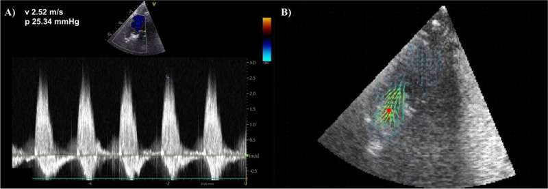
But the ongoing research of Ph.D. student Cameron Dockerill is looking into finding more accurate and non-invasive methods for assessing the pressure drop across the aortic valve. And his recent work confirms the overestimation of Doppler methods used in clinical practice, together with a promising alternative based on Blood Speckle Imaging (BSI).
Using a 3D printed model of an aortic valve that is placed inside a tube, with pressure sensors before and after, the team used cutting edge echocardiography probes to make BSI and Doppler acquisitions of blood flow across the valve.
BSI accurately estimated pressure drops up to 10.5 mmHg, which may be useful for estimations of trans-mitral valve and intra-cardiac pressure drops, but underestimated all of the greater pressure drops tested. Doppler overestimated pressure drop values of clinical significance, confirming its inaccuracies.
The key for the future is to capture the complete velocity profile of the blood traveling across the valve, which Doppler cannot do, before using the adequate formulation of the physical principles to convert the full velocity profile into a pressure drop more accurately.
"Our work has highlighted the limitations of existing non-invasive techniques used for the diagnosis and stratification of aortic stenosis patients. We are developing new techniques that are safer and more accurate, which we hope will improve disease diagnosis and patient outcomes in the future," says Cameron Dockerill, Ph.D. student, School of Biomedical Engineering & Imaging Sciences.
The study was published in Cardiovascular Ultrasound.
More information: Cameron Dockerill et al, Blood speckle imaging compared with conventional Doppler ultrasound for transvalvular pressure drop estimation in an aortic flow phantom, Cardiovascular Ultrasound (2022). DOI: 10.1186/s12947-022-00286-1


