Researchers identify genes that maintain healthy neurons
Researchers identify genes that maintain healthy neurons
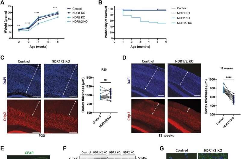
Dual loss of NDR kinases in neurons leads to neurodegeneration. (A) Line graph showing the average weight of NDR1 KO, NDR2 KO, NDR1/2 KO and control mice up to 6 wk of age. The differences in weight between the genotype groups at each time-point were analyzed using ordinary one-way ANOVAs or Kruskal–Wallis tests. n = 20–30 mice/group. (B) Graph illustrating the probability of survival of NDR1 KO, NDR2 KO, NDR1/2 KO and control mice up to 6 mo of age. n = 20–30 mice/group. (C, D) Immunofluorescence staining of Ctip2 in brain slices of NDR1/2 KO and control mice at P20 or 12 wk of age. White arrowed lines in the DAPI images show the thickness of the cortex, and white arrowed lines in the Ctip2 images show the thickness of the upper layers I–IV of the cortex. Scale bars: 200 μm. The graphs show quantifications of cortex thickness, and the data were analyzed using paired t tests. n = 18 measurements from three mice/genotype. (E) Immunofluorescence staining of GFAP in brain slices of 12-wk-old NDR1/2 KO and control mice. White arrows show areas with increased GFAP signal in NDR1/2 KO mice. (F) Western blot analyses of GFAP levels in lysates from the cortex of 6-wk-old mice. GAPDH was used as a loading control. The graphs show quantifications of the GFAP bands normalized against the GAPDH levels, and the data were analyzed using an ordinary one-way ANOVA with Tukey’s post hoc test. n = 3–5 mice/group. (G) Immunofluorescence staining of GFAP and the microglial marker Iba1 in the CA1 area of the hippocampus. White arrows indicate cells expressing the above-mentioned markers. Scale bars: 50 µm. (H) Images from brain slices of 12-wk-old Thy1-YFP–expressing mice in the CA1 area of the hippocampus. White arrowed lines indicate the stratum radiatum, where CA1 neuron dendrites are visible in YFP. The graph shows quantification of the hippocampal thickness (marked by the dashed yellow line—including stratum oriens, the CA1 cell body area, stratum radiatum and stratum lacunosum-moleculare), and the data were analyzed using a paired t test. n = 18 measurements from three mice/genotype. (I) Images from brain slices of Thy1-YFP–expressing mice in the CA1 cell body layer. The white arrow shows membrane protrusions present in NDR1/2 knockout neurons. Credit: Life Science Alliance (2022). DOI: 10.26508/lsa.202201712

Scientists at the Francis Crick Institute have found that deleting two genes that encode key enzyme proteins (kinases NDR1 and NDR2), impairs the health of neurons and leads to neurodegeneration in young mice as well as in adults.

Their study of mouse neurons highlights the essential role of these proteins in maintaining and preventing disease, a finding that could help with the discovery of future treatments for neurodegenerative diseases like Parkinson's and Amyotrophic lateral sclerosis (ALS).

As part of their work published in Life Science Alliance on November 29, the researchers set out to understand the role of kinase enzymes in the development of the nervous system and maintaining healthy neurons. For the first time in mice, they deleted genes that encode for both kinases NDR1 and NDR2 in neurons.

They found that deleting of either of the enzymes alone had no effect on neuronal health, but when both were eliminated simultaneously, the loss caused .

In order to understand why neurodegeneration occurs in the absence of these enzymes, the team further analyzed the and found accumulation of protein clusters tagged for removal, a key characteristic of many . This suggests that the kinase enzymes are essential for the neurons to perform autophagy, the process of removing old or damaged components.

"The neuron's ability to remove toxic proteins is an essential defense against neurodegeneration," says Sila Utanir, head of the Crick's Kinases and Brain Development Laboratory.

"Understanding that kinases NDR1 and NDR2 are vital to autophagy is important because if there was a way to boost their activity with future medicines, it could help clear the protein accumulation associated with disease."

The team also looked in even more detail at the mechanisms at play when these key enzymes are lost. They found that ATG9A, a protein found in some cellular membranes, which is associated with autophagy and lipid recycling, was incorrectly positioned and as such, couldn't function properly.

Flavia Roșianu, first author of the paper, said, "The complex signals sent between cells in our brain are all part of a bigger picture of neuronal health.

"In order to understand how our brain develops and why disease occurs, we need to piece together these connections and identify the most significant proteins and signals."

More information: Flavia Roşianu et al, Loss of NDR1/2 kinases impairs endomembrane trafficking and autophagy leading to neurodegeneration, Life Science Alliance (2022). DOI: 10.26508/lsa.202201712

Citation: Researchers identify genes that maintain healthy neurons (2022, November 30) retrieved 19 April 2024 from https://medicalxpress.com/news/2022-11-genes-healthy-neurons.html
This document is subject to copyright. Apart from any fair dealing for the purpose of private study or research, no part may be reproduced without the written permission. The content is provided for information purposes only.

Explore further

Enzyme pair play crucial role in maintaining balance of cellular processes

4 shares

Feedback to editors