This article has been reviewed according to Science X's editorial process and policies. Editors have highlighted the following attributes while ensuring the content's credibility:
fact-checked
peer-reviewed publication
trusted source
proofread
Investigating the interface between biomaterials and cells to help regenerate body tissues

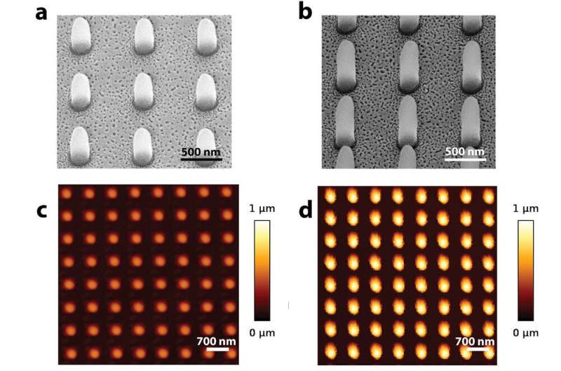
One of the approaches to improve the regeneration of body tissues is to focus on the physical surface cues of the biomaterial to see how different surface topographies such as tiny pillars may instruct cells "to do what we want them to do"—in this case regenerate bone tissue.
"The challenge is to create patterns on the surface that would support adhesion and differentiation of progenitor cells to become bone cells, but at the same time, discourage bacterial cells to grow, thereby preventing implant associated infections. So you structure the surface in such a way that bone cells like it but bacteria don't—that was a very clever idea which fascinated me," says Murali Ghatkesar of TU Delft.
"We are working on biomaterials that may help regenerate certain tissues for patients suffering from chronic diseases such as osteoarthritis and liver diseases," says collaborator Lidy Fratila-Apachitei (BMechE).
Fratila-Apachitei and Ghatkesar (PME) investigate how 3D printed pillars with features in the sub-micrometer range can affect adhesion and mechanics of living progenitor cells at single cell level. Together with postdoc researcher Livia Angeloni and the other collaborators, they developed and applied a novel method based on fluid force microscopy.
"By using our method, we could reveal early potential biophysical markers (e.g., cell adhesion strength) for osteogenic differentiation and matrix mineralization, and show that just a tiny difference in the height of the pillars of 0.5 micrometer is enough to change cell behavior," say the researchers.
The method and the results obtained enable a more rational design of physical surface cues for orthopedic biomaterials. Their results have been published in the journal Small and are featured on the inside front cover of the journal.
More information: Livia Angeloni et al, Fluidic Force Microscopy and Atomic Force Microscopy Unveil New Insights into the Interactions of Preosteoblasts with 3D‐Printed Submicron Patterns, Small (2022). DOI: 10.1002/smll.202204662


